. 2019 Jun 28;10(1):2885.
doi: 10.1038/s41467-019-10745-5.

Structural basis of TFIIH activation for nucleotide excision repair

Affiliations

Structural basis of TFIIH activation for nucleotide excision repair

Goran Kokic et al. Nat Commun. .

Abstract

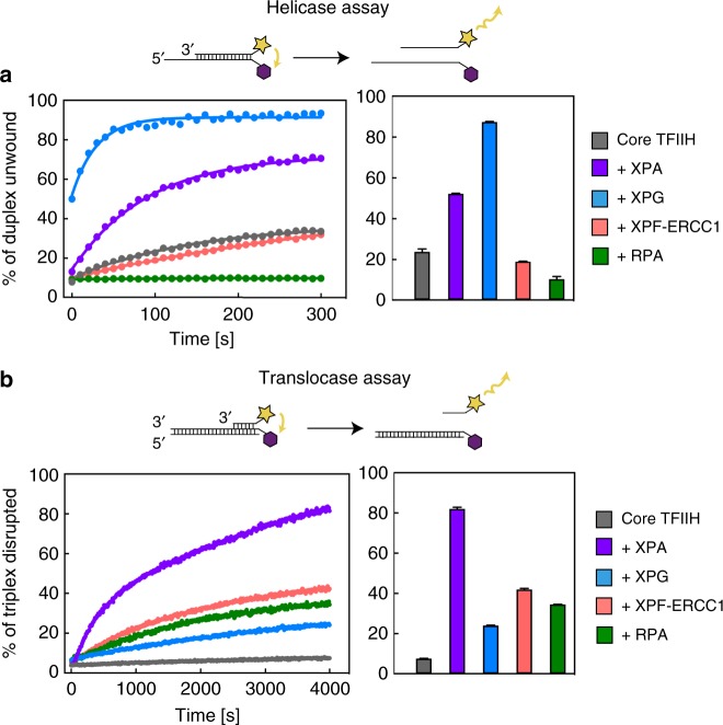
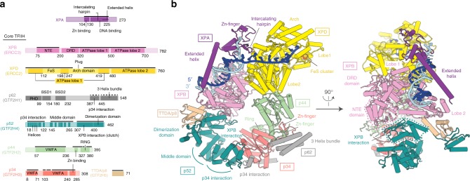
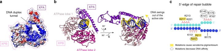
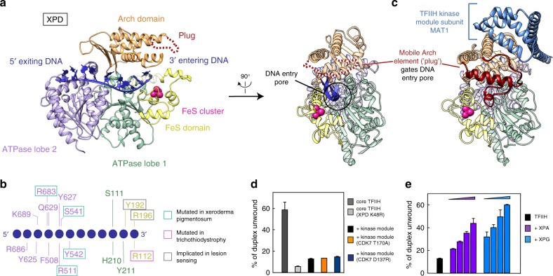
Nucleotide excision repair (NER) is the major DNA repair pathway that removes UV-induced and bulky DNA lesions. There is currently no structure of NER intermediates, which form around the large multisubunit transcription factor IIH (TFIIH). Here we report the cryo-EM structure of an NER intermediate containing TFIIH and the NER factor XPA. Compared to its transcription conformation, the TFIIH structure is rearranged such that its ATPase subunits XPB and XPD bind double- and single-stranded DNA, consistent with their translocase and helicase activities, respectively. XPA releases the inhibitory kinase module of TFIIH, displaces a 'plug' element from the DNA-binding pore in XPD, and together with the NER factor XPG stimulates XPD activity. Our results explain how TFIIH is switched from a transcription to a repair factor, and provide the basis for a mechanistic analysis of the NER pathway.


Conflict of interest statement

The authors declare no competing interests.

Figures

Fig. 1
Fig. 1
Regulation of ATPases in core transcription factor IIH. a Effect of nucleotide excision repair (NER) proteins on XPD 5′–3′ helicase activity. Real-time fluorescence measurement using a fluorescence energy transfer-based assay. Bars show the percentage of unwound product after 100 s (n = 2, error bars indicate the range of the data). RPA inhibits XPD helicase activity, by masking the single-stranded DNA overhang. Source Data are provided in the Source Data file. b Effect of NER proteins on XPB translocase activity. Real-time fluorescence measurement of triplex disruption. Bars show percentage of disrupted triplex after 4000 s (n = 2, error bars indicate the range of the data). Source Data are provided in the Source Data file
Fig. 2
Fig. 2
Structure of human core transcription factor IIH (TFIIH)-XPA-DNA complex. a Domain organization of XPA and human TFIIH subunits. Residues at domain borders are indicated. Solid and dashed black lines mark residues modeled as atomic and backbone structures, respectively. DRD damage recognition domain, NTE N-terminal extension, BSD BTF2-like, synapse-associated and DOS2-like domains, VWFA von Willebrand factor type A domain. b Cylindrical representation of the structure. Proteins colored as in a. Newly built XPB and p52 domains are highlighted with dashed lines and reveal how XPB is connected to the TFIIH core
Fig. 3
Fig. 3
XPA–DNA interactions. a DNA duplex tunnel formed by XPA and XPB. Blue, white, and red color indicates positive, neutral, and negative electrostatic surface potential, respectively. Created with UCSF Chimera. b Two positions of DNA in the tunnel. Tightly bound DNA is in blue, dissociated DNA in yellow, ATPase lobe 1 of XPB in pink, ATPase lobe 2 in hot pink, and XPA in purple. c Electrostatic interactions between XPA and the DNA junction. DNA nucleotides are indicated as circles. Patches of positively charged residues in proximity to the DNA backbone are indicated. Residues that are mutated in Xeroderma pigmentosum are highlighted in yellow. Mutation of encircled residues decreases DNA affinity
Fig. 4
Fig. 4
XPD activation and DNA binding. a Two views of XPD bound to DNA. XPD domains ATPase lobe 1, FeS, Arch, and lobe 2 are in green, yellow, orange, and medium purple, respectively. DNA is dark blue. A black circle depicts the DNA pore. b Schematic representation of XPD–DNA interactions. c Side view of XPD bound to the kinase module (PDB code 5OF4). The plug in the Arch domain is in dark red, the kinase module subunit MAT1 in blue. d Effect of kinase module variants on XPD helicase activity. Core transcription factor IIH (TFIIH) was incubated with two-fold excess of the kinase module and helicase activity was monitored as in Fig. 1a. Bars show the percentage of unwound product after 300 s (n = 2, error bars indicate the range of the data). Source Data are provided in the Source Data file. e Effect of increasing concentrations of XPA and XPG on XPD helicase activity in the presence of kinase module (0.25 μM core TFIIH, 0.5 μM kinase module, 0.375, 0.75, 1.5, or 3 μM XPA or XPG). Otherwise as in d. Source Data are provided in the Source Data file

References

    1. Scharer O. D. Nucleotide Excision Repair in Eukaryotes. Cold Spring Harbor Perspectives in Biology. 2013;5(10):a012609–a012609. doi: 10.1101/cshperspect.a012609. - DOI - PMC - PubMed
    1. Kraemer KH, et al. Xeroderma pigmentosum, trichothiodystrophy and Cockayne syndrome: a complex genotype-phenotype relationship. Neuroscience. 2007;145:1388–1396. doi: 10.1016/j.neuroscience.2006.12.020. - DOI - PMC - PubMed
    1. Lehmann AR. The xeroderma pigmentosum group D (XPD) gene: one gene, two functions, three diseases. Genes Dev. 2001;15:15–23. doi: 10.1101/gad.859501. - DOI - PubMed
    1. Evans E, Moggs JG, Hwang JR, Egly JM, Wood RD. Mechanism of open complex and dual incision formation by human nucleotide excision repair factors. EMBO J. 1997;16:6559–6573. doi: 10.1093/emboj/16.21.6559. - DOI - PMC - PubMed
    1. Compe E, Egly JM. Nucleotide excision repair and transcriptional regulation: TFIIH and beyond. Annu. Rev. Biochem. 2016;85:265–290. doi: 10.1146/annurev-biochem-060815-014857. - DOI - PubMed

Publication types

MeSH terms