

doi: 10.1038/s41598-019-45782-z.
Raman scattering yields cubic crystal grain orientation
Affiliations
- PMID: 31253866
- PMCID: PMC6599203
- DOI: 10.1038/s41598-019-45782-z
Item in Clipboard
Raman scattering yields cubic crystal grain orientation
Sci Rep.
.
Abstract
The paper proposes a fully optical method for determination of a cubic crystal grain orientation in a sample inspected by a Raman microscope. The method is based on a universal and strong polarisation anisotropy of the Raman scattering by doubly degenerate optic phonon modes and it only requires a standard Raman microscope equipped with a polarisation analysis. Explicit formulas for the orientation of the crystal grain are derived. The feasibility of the approach is demonstrated by comparing grain orientations in a polycrystalline cubic lacunar spinel GaV4S8 determined independently using electron backscatter diffraction and Raman scattering methods.
Conflict of interest statement
The authors declare no competing interests.
Figures
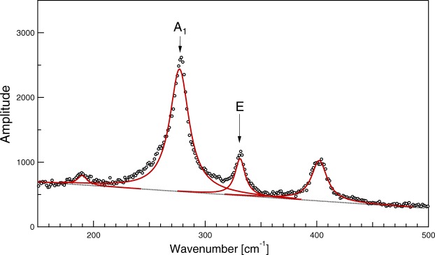
Typical parallel-polarised Raman spectrum detected from an arbitrarily oriented grain.
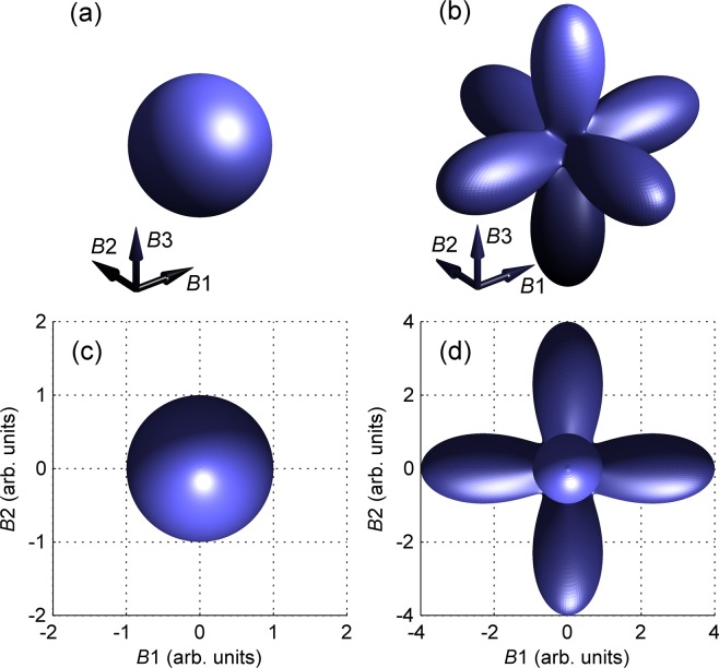
Polar plot of the polarisation-dependent factor of Raman scattering by (a) A1 and (b) E-symmetry modes in a cubic crystal calculated using equations (1, 2, 4 and 5) for a = b = 1. B1 − B3 stand for the three perpendicular directions parallel to the fourfold symmetry axes of the crystal. Projections along the B3 axis are shown in (c and d) panels, respectively.
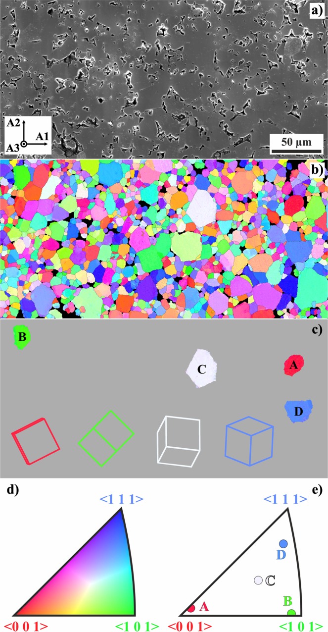
Grain morphology of the investigated GaV4S8 ceramics. (a) Scanning electron microscope image of an area of interest with sample coordinate system as an inset; (b) Inverse pole figure map from EBSD analysis of the area in (a); (c) positions of four grains chosen for detailed Raman measurements (, , , ), indicated projections of elementary cubic cells clarify their crystallographic orientation; (d) unit triangle with the color code of the inverse pole figure map; (e) surface normals of the chosen grains as determined from EBSD.
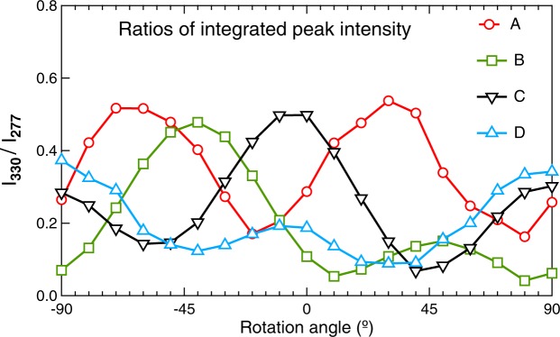
Parallel-polarised Raman scattering intensity ratios detected from the grain A (circles), B (squares) and C (triangles with vertex down) and D (triangles with vertex up) at selected frequencies as a function of the angle between the polariser and the reference direction A1 on the sample surface (indicated in Fig. 3). The intensity ratios are obtained from the raw measured data after the flat background subtraction as the integrated intensity at 330 ± 5 cm−1, divided by the integrated intensity at 277 ± 5 cm−1, I330/I277.
References
-
- Hlinka J, Pokorny J, Karimi S, Reaney IM. Angular dispersion of oblique phonon modes in BiFeO3 from micro-Raman scattering. Phys. Rev. B. 2011;83:020101. doi: 10.1103/PhysRevB.83.020101. - DOI
-
- Borodavka F, Pokorny J, Hlinka J. Combined piezoresponse force microscopy and Raman scattering investigation of domain boundaries in BiFeO3 ceramics. Phase Transitions. 2016;89:746–751. doi: 10.1080/01411594.2016.1206544. - DOI
LinkOut - more resources
Full Text Sources
