. 2019 Jul;51(7):1123-1130.
doi: 10.1038/s41588-019-0440-9. Epub 2019 Jun 28.

The genetic evolution of metastatic uveal melanoma

Affiliations

The genetic evolution of metastatic uveal melanoma

A Hunter Shain et al. Nat Genet. 2019 Jul.

Abstract

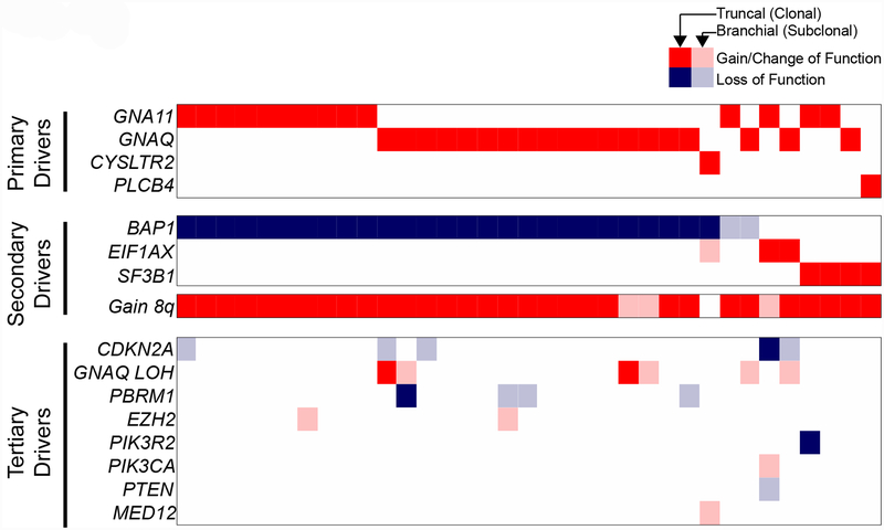
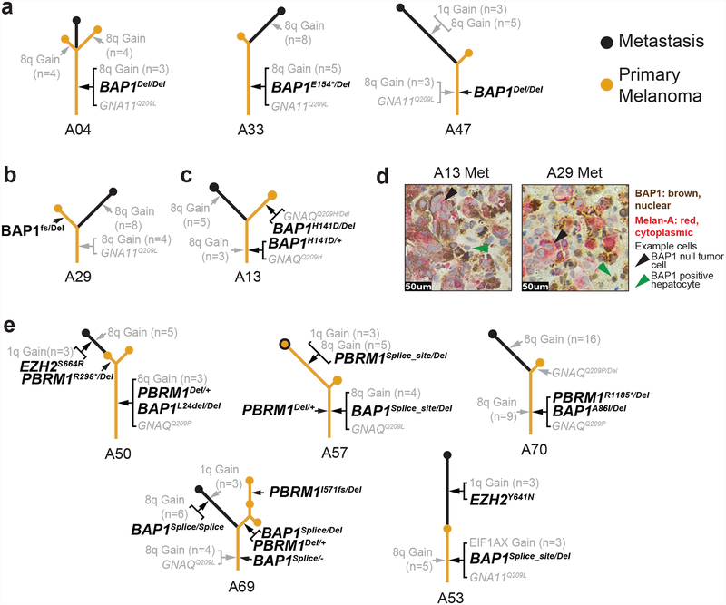
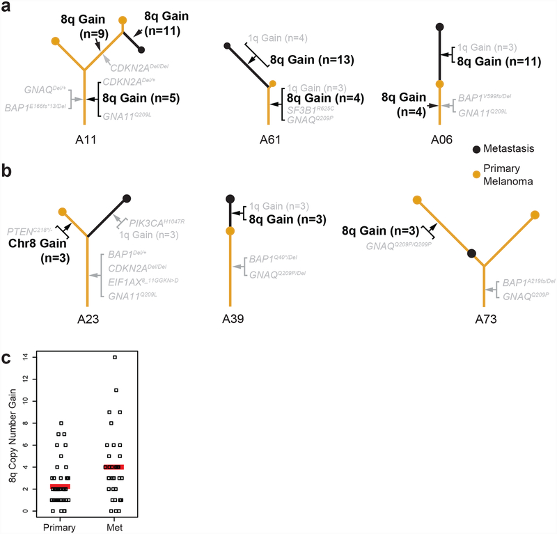
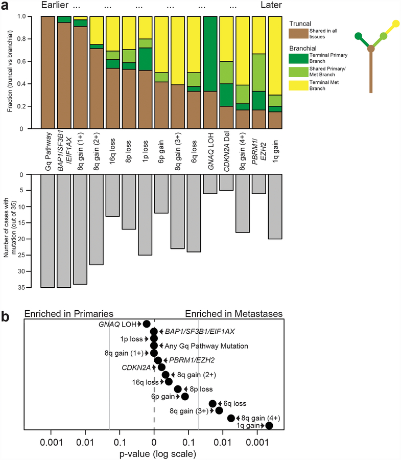
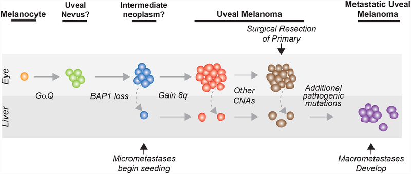
Uveal melanoma is a clinically distinct and particularly lethal subtype of melanoma originating from melanocytes in the eye. Here, we performed multi-region DNA sequencing of primary uveal melanomas and their matched metastases from 35 patients. We observed previously unknown driver mutations and established the order in which these and known driver mutations undergo selection. Metastases had genomic alterations distinct from their primary tumors; metastatic dissemination sometimes occurred early during the development of the primary tumor. Our study offers new insights into the genetics and evolution of this melanoma subtype, providing potential biomarkers for progression and therapy.


Conflict of interest statement

Competing Interests

B.C.B. is a consultant for Lilly Inc.

Figures

Figure 1 |
Figure 1 |. The patterns of somatic alterations within a patient’s tumor areas provide insights in the sequential order in which they arose (example case A11)
a, H&E sections of the enucleation specimen show morphologically distinct areas. The metastatic tissue stemmed from a liver core biopsy of the patient. Dotted regions were microdissected for genomic analyses. b, Point mutations are stratified by their mutant allele fractions (MAFs) in the two regions of the primary tumor and the metastasis. The blue bars indicate the expected MAFs of fully-clonal, heterozygous mutations after accounting for stromal cell contamination, with shadings indicating confidence intervals where those mutations should reside. Note the presence of a GNA11Q209L mutation that is fully clonal and heterozygous in all samples. By contrast, the BAP1Y173C mutation has an elevated MAF, indicating that it underwent loss-of-heterozygosity. c, Heatmap displaying copy number increases (red) or decreases (blue) for each sample (rows) across the genome with chromosomal boundaries indicated. Additional details related to deletion of CDKN2A and gain of chromosomal arm 8q are shown in Supplementary Figure 1. d, Inferred evolution of case A11 with mutations shared among all samples forming the trunk, and mutations not present in all samples forming the branches. The length of the trunk and branches is scaled by the total number of mutations in the samples.
Figure 2 |
Figure 2 |. The spectrum of driver mutations in uveal melanoma.
Multi-region sequencing of primary and matched metastases was performed on tumors from 35 patients. The spectrum of driver mutations (rows) in all patients (columns) is shown. Gain/change-of-function and loss-of-function mutations are respectively indicated as red or blue tiles. Mutations present in every sequenced region from a given patient are portrayed by solid tiles, and mutations present in only a subset of tissues from a given patient are portrayed by faded tiles. Mutations are grouped into three categories based on their deduced order of emergence during progression: primary drivers (shared and fully clonal), secondary drivers (shared and usually clonal), and tertiary drivers (private to later stages and typically subclonal).
Figure 3 |
Figure 3 |. Mutations disrupting chromatin remodeling factors emerge throughout the progression to metastatic disease.
a–c, Exemplary phylogenetic trees in which BAP1 loss emerged early (a) and late (b,c). In cases with early loss, bi-allelic loss-of-function mutations reside on the trunks of the trees, whereas in cases where it arose later, at least one hit is situated on a branch. d, Dual immunostaining for BAP1 (brown) and melan-A (red) demonstrates that A13 and A29 metastases are null or BAP1 protein. e, Cases in which mutations in other chromatin remodeling factors were detected. These alterations tended to arise later, positioning them on the branches.
Figure 4 |
Figure 4 |. Gene dosage of chromosome 8q increases during progression.
a, Examples of phylogenetic trees in which gains of 8q occur early and subsequently increase (see Supplementary Figs. 1b and 4 for more details on these copy number alterations). b, Examples of phylogenetic trees of tumors in which metastatic dissemination preceded gain of 8q. The detailed evolution of each case is available in the supplementary dataset (see Methods). c, Copy number increase of 8q in metastases and primary tumors is shown on the y-axis, with 0 reflecting the normal, diploid state. Red bars denote averages, indicating that in our cohort the level of 8q copy number increase doubled from primaries to metastases (P = 0.002, two-tailed t-test).
Figure 5 |
Figure 5 |. The sequential order of genetic alterations during metastatic progression.
a, Top panel: the y-axis indicates the relative fraction of patients in which the indicated somatic alterations occupied the trunk or branches of their respective phylogenetic trees. Somatic alterations that were more common in the trunks of phylogenetic trees were assumed to undergo selection earlier than those situated on their branches. Bottom panel: the number of patients with each of the indicated somatic alterations. b, Enrichment scores (P-values) were calculated using a two-tailed binomial test (see Methods for details) for the 15 genetic alterations highlighted in a to determine whether they were more common in primary tumors (n = 53) or in metastases (n = 36). Vertical gray lines indicate P-values of 0.05. Scores were calculated under the assumption (null hypothesis) that somatic alterations are equally distributed between primaries and metastases. Most somatic alterations were enriched in metastases, though only 6q loss, gain of at least 3 copies of 8q, and gain of 1q reached statistical significance.
Figure 6 |
Figure 6 |. The evolution of metastatic uveal melanoma.
‘CNA’, copy number alteration. ‘Other CNAs’ include combinations of 16q loss, 8p loss, 6q loss, or 6p gain, among others. ‘Additional pathogenic mutations’ include various combinations of 8q amplification, 1q gain, CDKN2A loss, SWI/SNF mutations, and EZH2 mutations, among others. The precursors to primary uveal melanoma are not well understood, as reflected here by the questions marks adjacent to those clinical stages in our model.

References

    1. Bastian BC The molecular pathology of melanoma: an integrated taxonomy of melanocytic neoplasia. Annu. Rev. Pathol 9, 239–271 (2014). - PMC - PubMed
    1. Singh AD & Topham A Survival rates with uveal melanoma in the United States: 1973–1997. Ophthalmology 110, 962–965 (2003). - PubMed
    1. Singh AD, Turell ME & Topham AK Uveal melanoma: trends in incidence, treatment, and survival. Ophthalmology 118, 1881–1885 (2011). - PubMed
    1. Kujala E, Mäkitie T & Kivelä T Very long-term prognosis of patients with malignant uveal melanoma. Invest. Ophthalmol. Vis. Sci 44, 4651–4659 (2003). - PubMed
    1. Dogrusöz M et al. The prognostic value of AJCC staging in uveal melanoma is enhanced by adding chromosome 3 and 8q status. Invest. Ophthalmol. Vis. Sci 58, 833–842 (2017). - PubMed

Methods-only references

    1. Shain AH et al. The genetic evolution of melanoma from precursor lesions. N. Engl. J. Med 373, 1926–1936 (2015). - PubMed
    1. Shain AH et al. Exome sequencing of desmoplastic melanoma identifies recurrent NFKBIE promoter mutations and diverse activating mutations in the MAPK pathway. Nat. Genet 47, 1194–1199 (2015). - PMC - PubMed
    1. Shain H Compiled genetic summary of the evolution of 35 metastatic uveal melanomas (2019). doi:10.6084/m9.figshare.6845675.v1 - DOI
    1. Shain AH et al. Genomic and transcriptomic analysis reveals incremental disruption of key signaling pathways during melanoma evolution. Cancer Cell 34, 45–55.e4 (2018). - PMC - PubMed
    1. Schouten JP et al. Relative quantification of 40 nucleic acid sequences by multiplex ligation-dependent probe amplification. Nucleic Acids Res 30, e57 (2002). - PMC - PubMed

Publication types

Substances