Review
. 2019 Dec;76(23):4589-4611.
doi: 10.1007/s00018-019-03203-4. Epub 2019 Jun 28.

Mechanisms of PINK1, ubiquitin and Parkin interactions in mitochondrial quality control and beyond

Affiliations
Review

Mechanisms of PINK1, ubiquitin and Parkin interactions in mitochondrial quality control and beyond

Andrew N Bayne et al. Cell Mol Life Sci. 2019 Dec.

Abstract

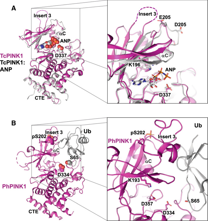
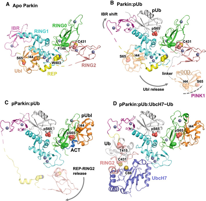
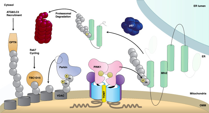
Parkinson's disease (PD) is a degenerative movement disorder resulting from the loss of specific neuron types in the midbrain. Early environmental and pathophysiological studies implicated mitochondrial damage and protein aggregation as the main causes of PD. These findings are now vindicated by the characterization of more than 20 genes implicated in rare familial forms of the disease. In particular, two proteins encoded by the Parkin and PINK1 genes, whose mutations cause early-onset autosomal recessive PD, function together in a mitochondrial quality control pathway. In this review, we will describe recent development in our understanding of their mechanisms of action, structure, and function. We explain how PINK1 acts as a mitochondrial damage sensor via the regulated proteolysis of its N-terminus and the phosphorylation of ubiquitin tethered to outer mitochondrial membrane proteins. In turn, phospho-ubiquitin recruits and activates Parkin via conformational changes that increase its ubiquitin ligase activity. We then describe how the formation of polyubiquitin chains on mitochondria triggers the recruitment of the autophagy machinery or the formation of mitochondria-derived vesicles. Finally, we discuss the evidence for the involvement of these mechanisms in physiological processes such as immunity and inflammation, as well as the links to other PD genes.

Keywords: Kinase; Mitochondria; PINK1; Parkin; Parkinson; Ubiquitin.


Conflict of interest statement

ANB declares no conflicts of interest. J-FT is a consultant for Mitokinin Inc. and a founding member of M4ND Pharma Inc.

Figures

Fig. 1
Fig. 1
Schematic of PINK1 import and processing. a PINK1 import and its proteolytic cycle in healthy, polarized mitochondria. PINK1 is translated on cytosolic ribosomes and threaded through TOM/TIM into the matrix, where it is subjected to its proteolytic cascade. After being cleaved by MPP, then PARL, it is retro-translocated and degraded. OMA1 serves as a surveillance mechanism for PINK1 that may escape TOM arrest. 3E glutamic acid triad E112/E113/E117 which gate its TOM7 arrest, TMD the transmembrane domain in which lies the PARL cleavage site, OMS the outer mitochondrial localization signal, MTS mitochondrial targeting signal. b PINK1 accumulation on depolarized mitochondria. Following mitochondrial damage, PINK1 is arrested in the TOM complex. MTS passage into the matrix is disrupted, as represented by the transparent TIM complex. Due to this, the PINK1 N-terminus is unable to be cleaved by MPP or PARL, and its N-terminus is left unprocessed. PINK1 oligomerizes and phosphorylates its dimeric partner in trans. Phosphorylation is depicted by the gold circles. Arrows coming from PINK1 depict phosphorylation of the Parkin Ubl domain (left), Ub tethered to an OMM protein (right), or itself (center)
Fig. 2
Fig. 2
Structures of PINK1 in the apo-, nucleotide-, and Ub-bound forms. a Structures of TcPINK1 (pdb 5OAT, chain A, magenta) and TcPINK1 bound to AMP-PNP (pdb 5YJ9, white). The inset on the right shows the nucleotide-binding site. The invariant Lys196 interacts with the α, β-phosphate groups of AMP-PNP and the catalytic base (Asp337) is in proximity to the γ-phosphate group. Both proteins had phosphomimetic mutations at the main phosphorylation site (S205E and S205D). Insert 3 is disordered in both structures. b Structure of PhPINK1 (pdb 6EQI, chain C, magenta) in complex with the Ub T66V-L71N mutant (chain A, gray), which adopts the minor extended conformation. The structure was determined in complex with a single-chain antibody (chain B, not shown). The inset shows the features of the Ub-binding site. The phosphate group on Ser202 stabilizes insert 3, which is then primed to make interactions with Ub. The structure also reveals a dramatic 90° rotation of the αC helix. The extended conformation of Ub exposes the side chain of Ser65, making it available for deprotonation by the catalytic base Asp334
Fig. 3
Fig. 3
Structural model of Parkin activation by PINK1. a Structure of human Parkin in the apo, autoinhibited conformation (pdb 5C1Z, chain A). The domains colors are defined and maintained throughout the figure. Key residues such as the sites of activation by mutagenesis (Phe146, Trp403) are shown as spheres. Zinc atoms are shown as gray spheres. b Structure of human Parkin bound to pUb (pdb 5N2W). The binding of pUb induces a straightening of the last helix in RING1 and a movement of the IBR domain. This leads to the release of the Ubl domain (transparent cartoon), which exposes residues such as Ile44 that are essential for Ubl phosphorylation at Ser65 by PINK1. c Structure of human phospho-Parkin bound to pUb (pdb 6GLC). The structure shows pUbl and the ACT element bound to RING0 and competing with RING2, which is released along with the REP (transparent cartoon). d Structure of fly phospho-Parkin bound to pUb and UbcH7 (pdb 6DJW). The position of Ub tethered to UbcH7 (transparent gray) was modeled on the NMR-based model of Parkin bound to UbcH7~Ub (pdb 6N13). The RING2 domain was positioned such that the acceptor cysteine Cys431 is located near Cys86 in UbcH7, to which Ub is conjugated
Fig. 4
Fig. 4
Schematic of Parkin activation and substrate ubiquitination on mitochondria. Representation of an ER–mitochondria contact site summarizing Parkin activation, ubiquitination of substrates, and its downstream consequences. TOM-arrested PINK1 oligomerizes, then phosphorylates ubiquitin on the OMM-localized proteins Mfn2 (right) and VDAC (left). These pUb chains recruit cytosolic Parkin (left), which continues to amplify this signal by transferring Ub from its active site Cys431 (shown as a circle on Parkin), onto substrate lysines. Ubiquitinated Mfn2 (right) recruits p97, which extracts Mfn2 from the OMM and permits its proteasomal degradation. PolyUb-VDAC (left) recruits the autophagy receptor OPTN, which in turn will catalyze autophagosome formation via recruitment of ATG8/LC3 and initiate subsequent mitophagy. DiUb-VDAC (left) is shown to recruit TBC1D15 to initiate Rab7 cycling

References

    1. Spillantini MG, Schmidt ML, Lee VM, Trojanowski JQ, Jakes R, Goedert M. Alpha-synuclein in Lewy bodies. Nature. 1997;388:839–840. - PubMed
    1. Langston JW, Ballard PA., Jr Parkinson’s disease in a chemist working with 1-methyl-4-phenyl-1,2,5,6-tetrahydropyridine. N Engl J Med. 1983;309:310. - PubMed
    1. Langston JW, Ballard P, Tetrud JW, Irwin I. Chronic Parkinsonism in humans due to a product of meperidine-analog synthesis. Science. 1983;219:979–980. - PubMed
    1. Schapira AH, Cooper JM, Dexter D, Jenner P, Clark JB, Marsden CD. Mitochondrial complex I deficiency in Parkinson’s disease. Lancet. 1989;1:1269. - PubMed
    1. Bender A, Krishnan KJ, Morris CM, Taylor GA, Reeve AK, Perry RH, Jaros E, Hersheson JS, Betts J, Klopstock T, Taylor RW, Turnbull DM. High levels of mitochondrial DNA deletions in substantia nigra neurons in aging and Parkinson disease. Nat Genet. 2006;38:515–517. - PubMed

LinkOut - more resources