. 2019 Oct;138(4):535-550.
doi: 10.1007/s00401-019-02040-w. Epub 2019 Jun 26.

Evidence for bidirectional and trans-synaptic parasympathetic and sympathetic propagation of alpha-synuclein in rats

Affiliations

Evidence for bidirectional and trans-synaptic parasympathetic and sympathetic propagation of alpha-synuclein in rats

Nathalie Van Den Berge et al. Acta Neuropathol. 2019 Oct.

Abstract

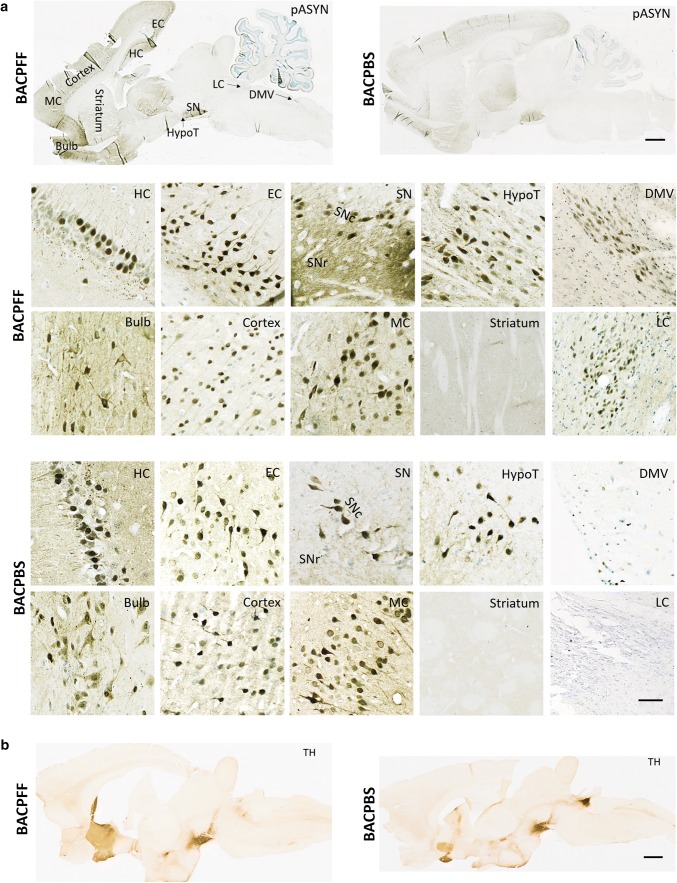
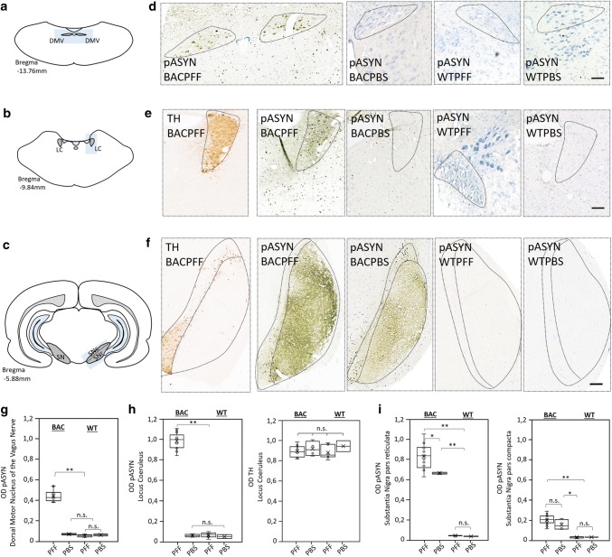
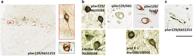
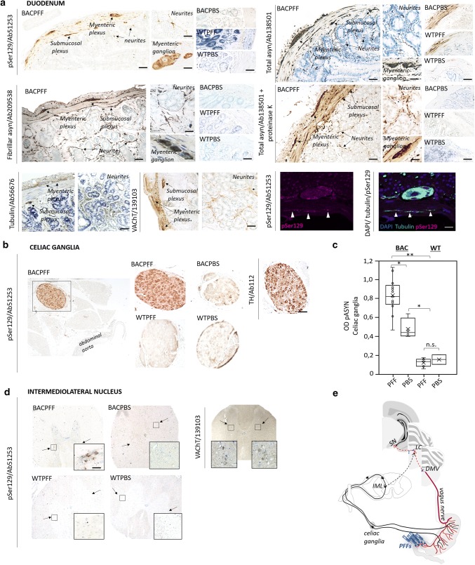
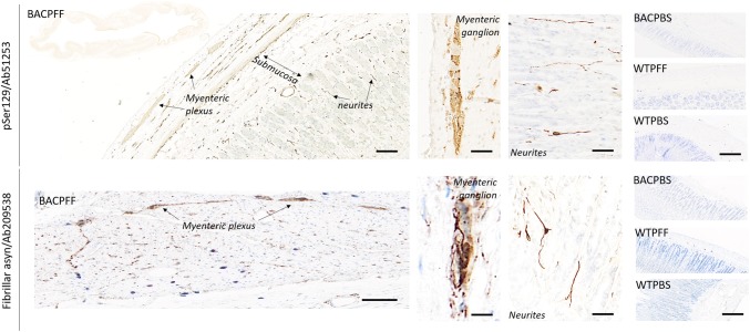
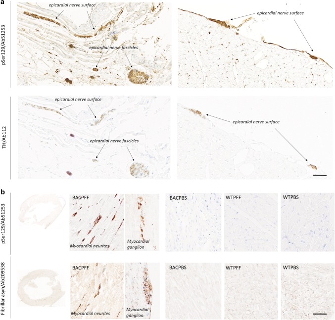
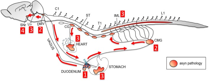
The conversion of endogenous alpha-synuclein (asyn) to pathological asyn-enriched aggregates is a hallmark of Parkinson's disease (PD). These inclusions can be detected in the central and enteric nervous system (ENS). Moreover, gastrointestinal symptoms can appear up to 20 years before the diagnosis of PD. The dual-hit hypothesis posits that pathological asyn aggregation starts in the ENS, and retrogradely spreads to the brain. In this study, we tested this hypothesis by directly injecting preformed asyn fibrils into the duodenum wall of wild-type rats and transgenic rats with excess levels of human asyn. We provide a meticulous characterization of the bacterial artificial chromosome (BAC) transgenic rat model with respect to initial propagation of pathological asyn along the parasympathetic and sympathetic pathways to the brainstem, by performing immunohistochemistry at early time points post-injection. Induced pathology was observed in all key structures along the sympathetic and parasympathetic pathways (ENS, autonomic ganglia, intermediolateral nucleus of the spinal cord (IML), heart, dorsal motor nucleus of the vagus, and locus coeruleus (LC)) and persisted for at least 4 months post-injection. In contrast, asyn propagation was not detected in wild-type rats, nor in vehicle-injected BAC rats. The presence of pathology in the IML, LC, and heart indicate trans-synaptic spread of the pathology. Additionally, the observed asyn inclusions in the stomach and heart may indicate secondary anterograde propagation after initial retrograde spreading. In summary, trans-synaptic propagation of asyn in the BAC rat model is fully compatible with the "body-first hypothesis" of PD etiopathogenesis. To our knowledge, this is the first animal model evidence of asyn propagation to the heart, and the first indication of bidirectional asyn propagation via the vagus nerve, i.e., duodenum-to-brainstem-to-stomach. The BAC rat model could be very valuable for detailed mechanistic studies of the dual-hit hypothesis, and for studies of disease modifying therapies targeting early pathology in the gastrointestinal tract.

Keywords: Alpha-synuclein; Autonomic nervous system; BAC rat model; Parkinson’s disease; Prion-like spread.


Conflict of interest statement

The author declares that they have no competing interests.

Figures

Fig. 1
Fig. 1
a Distribution of phosphorylated asyn (pASYN/Ab51253) pathology in sagittal brain sections of S129A PFF- and PBS-injected BAC rats at 4 months post-injection. The scale bar represents 1 mm in the whole brain sagittal sections (upper panel). In the lower panel, representative high-magnification photomicrographs of phosphorylated asyn (pASYN, Ab51253) pathology are shown in several brain areas of S129A PFF- and PBS-injected BAC rats: bulb, cortex, motor cortex (MC), striatum, hippocampus (HC), entorhinal cortex (EC), substantia nigra pars compacta (SNc), substantia nigra pars reticulata (SNr) hypothalamus (HypoT), dorsal motor nucleus of the vagus nerve (DMV), locus coeruleus (LC). The scale bar represents 100 µm in the DMV and LC, and 50 µm in all other brain areas. Similar levels of brain pathology were seen in the BAPFF and BACPBS rats, except for the brainstem (DMV, LC, and SNr), which contained significantly less or no pathology in the BACPBS rats. b Distribution of tyrosine hydroxylase in a sagittal brain section of BACPFF and BACPBS rats. Scale bar: 1 mm
Fig. 2
Fig. 2
Phosphorylated asyn (pASYN) pathology in the DMV, LC, and SN of S129A PFF- and PBS-injected BAC rats and WT controls at 4 months post-injection. a–c Schematic of DMV (a), LC (b), and SN (c) anatomy at bregma − 13.76 mm, − 9.84 mm and − 5.88 mm, respectively. d High magnification photomicrograph of asyn pathology (pASYN/Ab51253) in the DMV of PFF- and PBS-injected BAC and WT rats. Scale bar: 200 µm. e, f High-magnification photomicrograph of tyrosine hydroxylase and asyn pathology (pASYN/Ab51253) in the LC (e) and SN (f) of PFF- and PBS-injected BAC and WT rats. Scalebar: 200 µm. g Optical density (OD) measurements of pASYN pathology in the DMV of PFF- and PBS-injected BAC and WT rats. The regions of interest used for the analysis are indicated in d and in Online Resource Fig. 6. h Optical density measurements of pASYN pathology (left panel) and TH (right panel) in the LC of PFF- and PBS-injected BAC and WT rats. The regions of interest used for the analysis are indicated in e and in Online Resource Fig. 6. The BACPFF rats showed pathology in the DMV and LC at 2 and 4 months post-injection (DMV: p < 0.01, LC: p < 0.001). The DMV and LC of BACPBS and WT rats remained free of pathology. i Optical density measurements of pASYN pathology in the SN pars reticulata (left panel) and pars compacta (right panel) of PFF- and PBS-injected BAC and WT rats. The regions of interest for the analysis are indicated in f and in Online Resource Fig. 6. The BACPFF rats showed significantly more pathology in the SNr (p < 0.05) at 2 and 4 months post-injection, but not in the SNc. The SN of WT rats remained free of pathology. Each data point represents mean OD values. Horizontal bars: ± SE
Fig. 3
Fig. 3
Representative high magnification photomicrographs of phosphorylated asyn-positive inclusions detected in the DMV of seeded BAC rats. Inclusions were evident in neuronal cell bodies (a, b) and as elongated Lewy neurite-like structures (c). Depending on the antibodies used, inclusions (a/b, scale bar = 25 µm) and neurites (c, scale bar = 50 µm) had different appearances
Fig. 4
Fig. 4
a Distribution of asyn pathology in the duodenum of S129A PFF- and PBS -injected BAC rats and WT controls, at 4 months post-injection, detected with four different antibodies. Scale bar: 100 µm. Representative high magnification photomicrographs are shown of the myenteric ganglion and neurites in the BACPFF. The enteric nervous system remained pathology free in BACPBS and WT rats. Scale bar: 50 µm. For co-localization purposes, representative high magnification photomicrographs are shown of the distribution of VAChT, tubulin, and DAPI in the duodenum. Asyn pathology co-localized with DAPI, VAChT, and tubulin. Scale bar: 50 µm. b Distribution of asyn pathology in the celiac ganglia of S129A PFF- and PBS-injected BAC rats and WT controls, at 4 months post-injection (left panel). Distribution of tyrosine hydroxylase in the celiac ganglion of a BAC PFF rat (right panel). The TH distribution is similar across the four experimental groups. Scale bar: 200 µm. c Optical density (OD) measurements of pASYN pathology in the celiac ganglia of PFF- and PBS-injected BAC and WT rats. pASYN levels were significantly higher in the ganglia of BACPFF compared to BACPBS rats (p < 0.05), and were close to zero in the WT rats. d Distribution of asyn pathology in the IML of S129A PFF- and PBS-injected BAC rats and WT controls, at 2 or 4 months post-injection (left panel). For co-localization purposes, distribution of VAChT in the IML of a BAC PFF rat. The VAChT distribution is similar across the four experimental groups (right panel). High magnification photomicrographs of the IML are shown in the right bottom corner of each image. 4/13 BACPFF showed unilateral asyn pathology and 9/13 BACPFF showed bilateral asyn pathology in the IML after duodenal seeding. No pathology was observed in the IML of BACPBS and WT rats (see Table 2 and Online Resourse Fig. 9). Scale bar: 50 µm. e Schematic of the proposed retrograde parasympathetic (duodenum → DMV → LC) and sympathetic (duodenum → celiac ganglia → IML → LC) spreading routes of asyn pathology
Fig. 5
Fig. 5
Distribution of asyn pathology in the stomach of S129A PFF- and PBS-injected BAC rats and WT controls, at 4 months post-injection, detected with two different antibodies. Scale bar = 100 µm. Representative high magnification photomicrographs are shown of the myenteric ganglion and neurites in the BACPFF. The plexus and lumen of the BACPBS and WT rats remained pathology free. Scale bar: 50 µm
Fig. 6
Fig. 6
a Distribution of asyn pathology (pSer129/Ab51253; upper panel) and tyrosine hydroxylase (lower panel) in the heart of S129A PFF- and PBS-injected BAC rats and WT controls at 4 months post-injection. In the BACPFF rats, clear co-localization was seen between staining for TH and phosphorylated asyn pathology. Scale bar = 100 µm. b Distribution of asyn pathology in the myocardium of S129A PFF- and PBS-injected BAC rats and WT controls, detected with two different antibodies. Representative high magnification photomicrographs are shown of a myocardial ganglion and neurites in the BACPFF. The heart remained free of pathology in all BACPBS and WT rats. Scale bar: 50 µm
Fig. 7
Fig. 7
Schematic overview of the hypothesized trans-synaptic bidirectional propagation of asyn pathology through the autonomic nervous system to the brain after gastrointestinal seeding. First, seeding of the duodenum with fibrils induces aggregation of endogenous asyn in the duodenum. Second, the induced pathology retrogradely spreads along the parasympathetic pathway to the dorsal motor nucleus of the vagus (DMV) and along the sympathetic pathway to the celiac ganglia (CMG). Third, asyn pathology propagates trans-synaptically to the locus coeruleus (LC) and intermediolateral nucleus of the spinal cord (IML). Additionally, asyn pathology spreads anterogradely to the heart and to the stomach. Fourth, asyn pathology spreads to the substantia nigra pars reticulata (SNr)

Comment in

References

    1. Abbott RD, Petrovitch H, White LR, Masaki KH, Tanner CM, Curb JD, et al. Frequency of bowel movements and the future risk of Parkinson’s disease. Neurology. 2001;57:456–462. doi: 10.1212/WNL.57.3.456. - DOI - PubMed
    1. Abdelmotilib H, Maltbie T, Delic V, Liu Z, Hu X, Fraser KB, et al. α-Synuclein Fibril-induced inclusion spread in rats and mice correlates with dopaminergic neurodegeneration. Neurobiol Dis. 2017;105:84–98. doi: 10.1016/j.nbd.2017.05.014. - DOI - PMC - PubMed
    1. Adler CH, Beach TG. Neuropathological basis of nonmotor manifestations of Parkinson’s disease. Mov Disord. 2016;31:1114–1119. doi: 10.1002/mds.26605. - DOI - PMC - PubMed
    1. Ayers JI, Brooks MM, Rutherford NJ, Howard JK, Sorrentino ZA, Riffe CJ, et al. Robust central nervous system pathology in transgenic mice following peripheral injection of α-Synuclein Fibrils. J Virol. 2017;91:e02095-16. doi: 10.1128/JVI.02095-16. - DOI - PMC - PubMed
    1. Beach TG, Adler CH, Sue LI, Vedders L, Lue L, White Iii CL, et al. Multi-organ distribution of phosphorylated alpha-synuclein histopathology in subjects with Lewy body disorders. Acta Neuropathol. 2010;119:689–702. doi: 10.1007/s00401-010-0664-3. - DOI - PMC - PubMed

Publication types

MeSH terms

Substances