. 2019 Oct;20(5):449-459.
doi: 10.1007/s10162-019-00727-1. Epub 2019 Jun 28.

Osteoclasts Modulate Bone Erosion in Cholesteatoma via RANKL Signaling

Affiliations

Osteoclasts Modulate Bone Erosion in Cholesteatoma via RANKL Signaling

Ryusuke Imai et al. J Assoc Res Otolaryngol. 2019 Oct.

Abstract

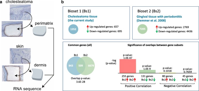
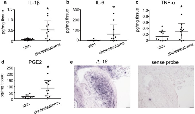
Cholesteatoma starts as a retraction of the tympanic membrane and expands into the middle ear, eroding the surrounding bone and causing hearing loss and other serious complications such as brain abscess and meningitis. Currently, the only effective treatment is complete surgical removal, but the recurrence rate is relatively high. In rheumatoid arthritis (RA), osteoclasts are known to be responsible for bone erosion and undergo differentiation and activation by receptor activator of NF-κB ligand (RANKL), which is secreted by synovial fibroblasts, T cells, and B cells. On the other hand, the mechanism of bone erosion in cholesteatoma is still controversial. In this study, we found that a significantly larger number of osteoclasts were observed on the eroded bone adjacent to cholesteatomas than in unaffected areas, and that fibroblasts in the cholesteatoma perimatrix expressed RANKL. We also investigated upstream transcription factors of RANKL using RNA sequencing results obtained via Ingenuity Pathways Analysis, a tool that identifies relevant targets in molecular biology systems. The concentrations of four candidate factors, namely interleukin-1β, interleukin-6, tumor necrosis factor α, and prostaglandin E2, were increased in cholesteatomas compared with normal skin. Furthermore, interleukin-1β was expressed in infiltrating inflammatory cells in the cholesteatoma perimatrix. This is the first report demonstrating that a larger-than-normal number of osteoclasts are present in cholesteatoma, and that the disease involves upregulation of factors related to osteoclast activation. Our study elucidates the molecular basis underlying bone erosion in cholesteatoma.

Keywords: IL-1β; RNA sequencing; cholesteatoma; fibroblast; osteoclast; receptor activator of NF-κB ligand (RANKL).


Conflict of interest statement

The authors declare that they have no conflicts of interest.

Figures

Fig. 1
Fig. 1
Elevated numbers of osteoclasts are present on the bone surface adjacent to cholesteatoma lesions. a Osteoclast cytoplasm is stained red using TRAP (scale bar: 10 μm). Multiple nuclei are stained blue. b Osteoclasts (arrows) are observed on the bone surface adjacent to cholesteatoma lesions (scale bar 50 μm). c The number of osteoclasts per μm2 on the bone surface adjacent to cholesteatoma lesions was larger than those on the surface of non-cholesteatomatous bone (N = 24). The asterisk indicates a statistically significant difference
Fig. 2
Fig. 2
Fibroblasts in the cholesteatoma perimatrix express RANKL. a Expression of RANKL mRNA in cholesteatoma, corrected for GAPDH mRNA, is significantly higher than in control skin (n = 9). The asterisk indicates a statistically significant difference. b DAB staining for RANKL in the cholesteatoma perimatrix, counterstained with hematoxylin (scale bars 50 μm). c Immunofluorescent staining of the cholesteatoma perimatrix. Co-localization of vimentin and RANKL immunofluorescence shows that fibroblasts in the perimatrix express RANKL. RANKL was labeled with Alexa Fluor® 555, and vimentin was labeled with Alexa Fluor® 488. Nuclei were stained with Hoechst® 33342 (scale bars: 10 μm)
Fig. 3
Fig. 3
Osteoclast differentiation is positively regulated in the cholesteatoma perimatrix. a Illustration of the technique used to dissect the perimatrix and dermis from the cholesteatoma and skin by laser microdissection. b Significance of the overlap between gene sets of cholesteatoma tissue and gingival tissue with periodontitis. The scale of the bars is measured in -log (P value). The accession number for the public data used for the graphs is GE16134
Fig. 4
Fig. 4
Concentrations of IL-1β, IL-6, TNF-α, and PGE2 in cholesteatomas. ad ELISA measurements show that the concentrations of IL-1β, IL-6, TNF-α, and PGE2 are higher in cholesteatoma than in skin (n = 11). The asterisks indicate statistically significant differences. e Mononuclear infiltrating cells in the cholesteatoma perimatrix express IL-1β mRNA, as shown in the left panel using in situ hybridization. The right panel shows in situ hybridization using a sense probe as a negative control (scale bars 50 μm)

References

    1. Abramson M, Moriyama H, Huang CC. Pathogenic factors in bone resorption in cholesteatoma. Acta Otolaryngol. 1984;97:437–442. doi: 10.3109/00016488409132918. - DOI - PubMed
    1. Akaogi J, Nozaki T, Satoh M, Yamada H. Role of PGE2 and EP receptors in the pathogenesis of rheumatoid arthritis and as a novel therapeutic strategy endocrine. Metab Immune Disord Drug Targets. 2006;6:383–394. doi: 10.2174/187153006779025711. - DOI - PubMed
    1. Ashburner M, Ball CA, Blake JA, Botstein D, Butler H, Cherry JM, Davis AP, Dolinski K, Dwight SS, Eppig JT, Harris MA, Hill DP, Issel-Tarver L, Kasarskis A, Lewis S, Matese JC, Richardson JE, Ringwald M, Rubin GM, Sherlock G. Gene ontology: tool for the unification of biology. The Gene Ontology Consortium. Nat Genet. 2000;25:25–29. doi: 10.1038/75556. - DOI - PMC - PubMed
    1. Boyle WJ, Simonet WS, Lacey DL. Osteoclast differentiation and activation. Nature. 2003;423:337–342. doi: 10.1038/nature01658. - DOI - PubMed
    1. Bresnihan B, et al. Treatment of rheumatoid arthritis with recombinant human interleukin-1 receptor antagonist. Arthritis Rheum. 1998;41:2196–2204. doi: 10.1002/1529-0131(199812)41:12<2196::Aid-art15>3.0.Co;2-2. - DOI - PubMed

Publication types