Review
. 2019 Oct;176(19):3741-3753.
doi: 10.1111/bph.14779. Epub 2019 Aug 9.

The role of smooth muscle cells in plaque stability: Therapeutic targeting potential

Affiliations
Review

The role of smooth muscle cells in plaque stability: Therapeutic targeting potential

Jennifer L Harman et al. Br J Pharmacol. 2019 Oct.

Abstract

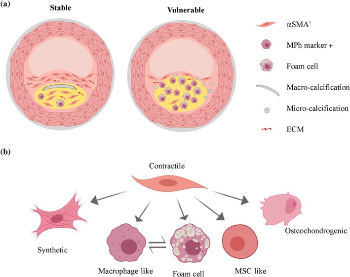
Events responsible for cardiovascular mortality and morbidity are predominantly caused by rupture of "vulnerable" atherosclerotic lesions. Vascular smooth muscle cells (VSMCs) play a key role in atherogenesis and have historically been considered beneficial for plaque stability. VSMCs constitute the main cellular component of the protective fibrous cap within lesions and are responsible for synthesising strength-giving extracellular matrix components. However, lineage-tracing experiments in mouse models of atherosclerosis have shown that, in addition to the fibrous cap, VSMCs also give rise to many of the cell types found within the plaque core. In particular, VSMCs generate a substantial fraction of lipid-laden foam cells, and VSMC-derived cells expressing markers of macrophages, osteochondrocyte, and mesenchymal stem cells have been observed within lesions. Here, we review recent studies that have changed our perspective on VSMC function in atherosclerosis and discuss how VSMCs could be targeted to increase plaque stability.


Conflict of interest statement

The authors declare no conflicts of interest.

Figures

Figure 1
Figure 1
Stable versus vulnerable atherosclerotic plaque and vascular smooth muscle cell (VSMC)‐derived plaque cell phenotypes. (a) A simplified scheme showing a stable lesion with a thick collagen‐rich (extracellular matrix [ECM]) fibrous cap covering a plaque core (yellow area), which contains a high ratio of αSMA‐positive (αSMA+) cells compared with cells expressing macrophage‐associated markers (MPh‐marker +) and macro‐calcified deposits. In contrast, vulnerable plaques have a thin fibrous cap, which often contains micro‐calcified deposits, fewer cells, and less ECM. The lipid‐rich core (yellow area) of vulnerable lesions includes numerous foam cells as well as a high ratio of cells expressing macrophage‐associated markers compared with αSMA‐positive cells. Please note that details such as endothelial cells, adventitial cells, and internal and external elastic lamina are not displayed. (b) Contractile VSMCs can alter their phenotype to a more active “synthetic” state in which they up‐regulate selective gene sets important for vascular remodelling, including cytokines, chemokines, proteases, and adhesion proteins. Within plaques, VSMCs also give rise to foam cells or express markers traditionally associated with other cell types, such as macrophages, mesenchymal stem cells (MSCs), or osteochondrocytes. The relative contribution of VSMC‐derived plaque cell phenotypes in stable versus vulnerable plaque remains unknown
Figure 2
Figure 2
Genetic lineage labelling of vascular smooth muscle cells (VSMCs). (a) Diagram of the Myh11–CreERt2 transgene and the ROSA26–YFP reporter allele. Tamoxifen‐induced recombination at the ROSA26–YFP locus results in the expression of YFP protein, which is stably propagated within progeny after high‐fat diet (HFD)‐induced atherogenesis independent of Myh11 expression levels (b) Schematic of the ROSA26–Confetti reporter allele. Tamoxifen‐induced recombination at the ROSA26–Confetti locus results in stochastic expression of one of four fluorescent proteins (nuclear GFP, YFP, RFP, or membrane‐associated CFP), which are stably propagated within progeny after HFD‐induced atherogenesis. Confocal images show non‐labelled and Myh11‐lineage Confetti‐labelled VSMCs in carotid arteries before and after HFD‐induced atherogenesis. Scale bars are 100 μm

References

    1. Ackers‐Johnson, M. , Talasila, A. , Sage, A. P. , Long, X. , Bot, I. , Morrell, N. W. , … Sinha, S. (2015). Myocardin regulates vascular smooth muscle cell inflammatory activation and disease. Arteriosclerosis, Thrombosis, and Vascular Biology, 35(4), 817–828. 10.1161/ATVBAHA.114.305218 - DOI - PMC - PubMed
    1. Adiguzel, E. , Ahmad, P. J. , Franco, C. , & Bendeck, M. P. (2009). Collagens in the progression and complications of atherosclerosis. Vascular Medicine (London, England), 14(1), 73–89. 10.1177/1358863X08094801 - DOI - PubMed
    1. Aikawa, E. , Nahrendorf, M. , Figueiredo, J. L. , Swirski, F. K. , Shtatland, T. , Kohler, R. H. , … Weissleder, R. (2007). Osteogenesis associates with inflammation in early‐stage atherosclerosis evaluated by molecular imaging in vivo. Circulation, 116(24), 2841–2850. 10.1161/CIRCULATIONAHA.107.732867 - DOI - PubMed
    1. Aikawa, M. , Sakomura, Y. , Ueda, M. , Kimura, K. , Manabe, I. , Ishiwata, S. , … Nagai, R. (1997). Redifferentiation of smooth muscle cells after coronary angioplasty determined via myosin heavy chain expression. Circulation, 96(1), 82–90. 10.1161/01.CIR.96.1.82 - DOI - PubMed
    1. Albarran‐Juarez, J. , Kaur, H. , Grimm, M. , Offermanns, S. , & Wettschureck, N. (2016). Lineage tracing of cells involved in atherosclerosis. Atherosclerosis, 251, 445–453. 10.1016/j.atherosclerosis.2016.06.012 - DOI - PubMed

Publication types

MeSH terms