. 2019 Sep;110(9):2806-2821.
doi: 10.1111/cas.14118. Epub 2019 Jul 23.

Establishment of a novel experimental model for muscle-invasive bladder cancer using a dog bladder cancer organoid culture

Affiliations

Establishment of a novel experimental model for muscle-invasive bladder cancer using a dog bladder cancer organoid culture

Mohamed Elbadawy et al. Cancer Sci. 2019 Sep.

Abstract

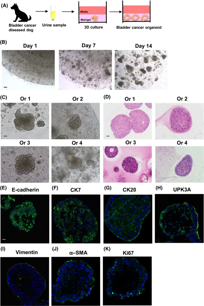
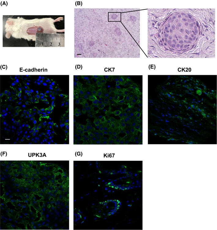
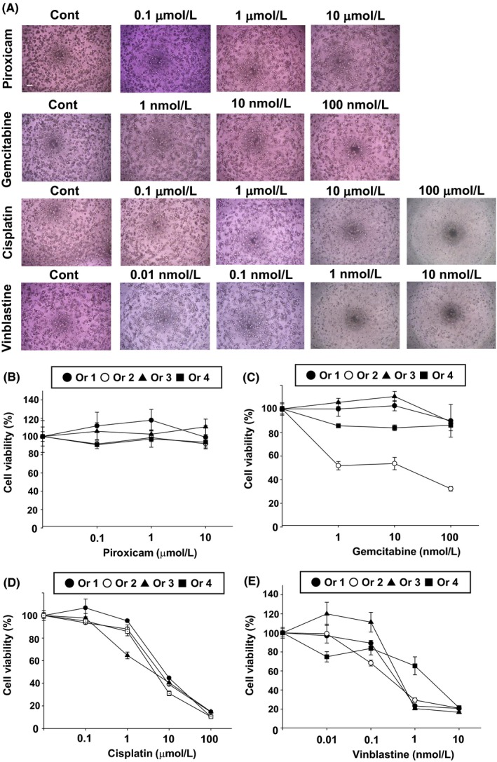
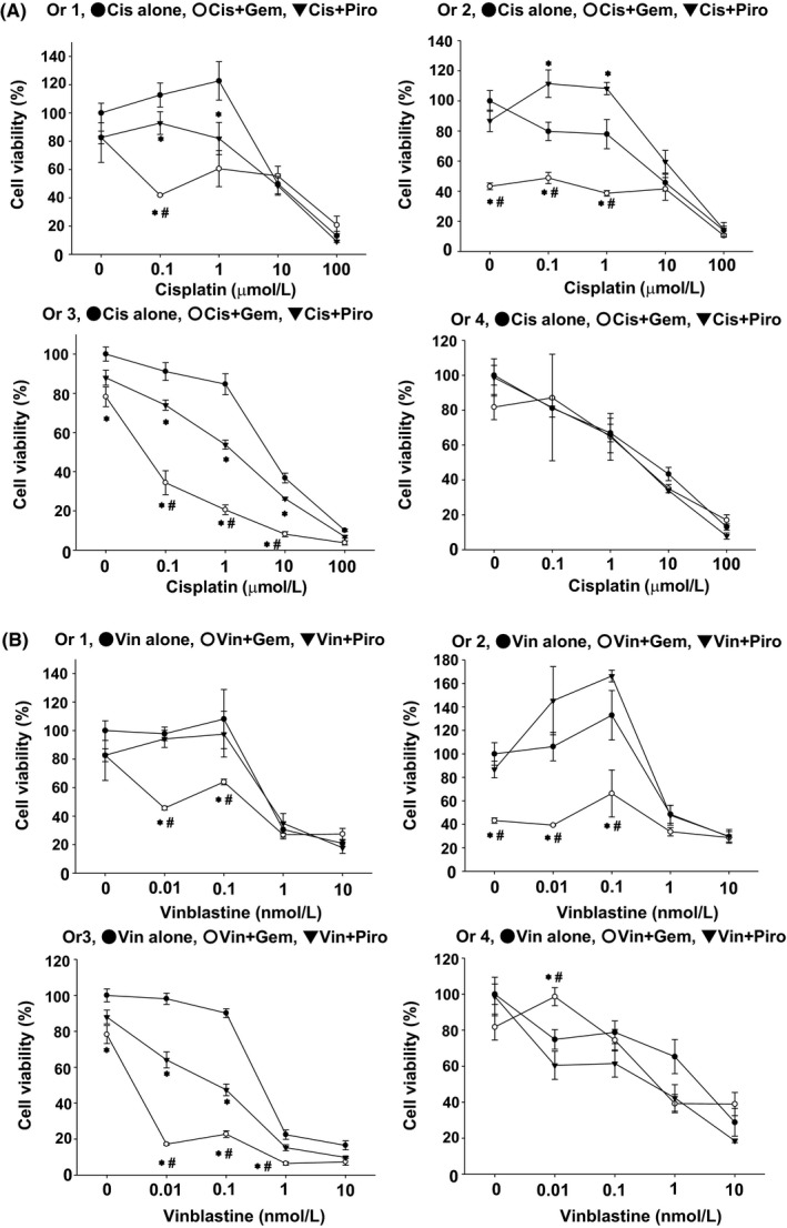
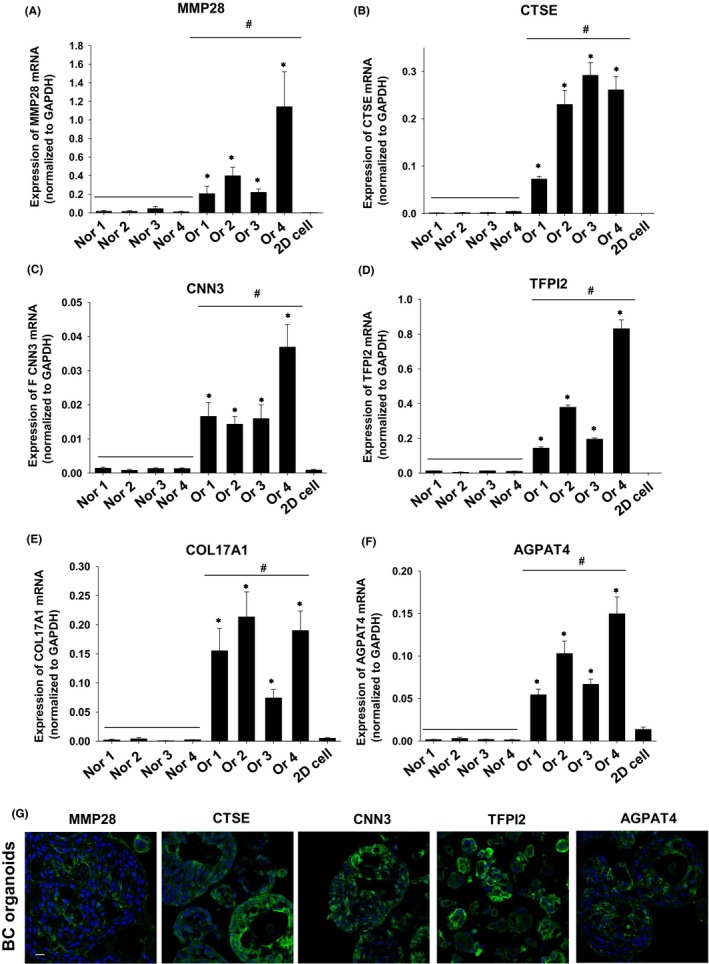
In human and dogs, bladder cancer (BC) is the most common neoplasm affecting the urinary tract. Dog BC resembles human muscle-invasive BC in histopathological characteristics and gene expression profiles, and could be an important research model for this disease. Cancer patient-derived organoid culture can recapitulate organ structures and maintains the gene expression profiles of original tumor tissues. In a previous study, we generated dog prostate cancer organoids using urine samples, however dog BC organoids had never been produced. Therefore we aimed to generate dog BC organoids using urine samples and check their histopathological characteristics, drug sensitivity, and gene expression profiles. Organoids from individual BC dogs were successfully generated, expressed urothelial cell markers (CK7, CK20, and UPK3A) and exhibited tumorigenesis in vivo. In a cell viability assay, the response to combined treatment with a range of anticancer drugs (cisplatin, vinblastine, gemcitabine or piroxicam) was markedly different in each BC organoid. In RNA-sequencing analysis, expression levels of basal cell markers (CK5 and DSG3) and several novel genes (MMP28, CTSE, CNN3, TFPI2, COL17A1, and AGPAT4) were upregulated in BC organoids compared with normal bladder tissues or two-dimensional (2D) BC cell lines. These established dog BC organoids might be a useful tool, not only to determine suitable chemotherapy for BC diseased dogs but also to identify novel biomarkers in human muscle-invasive BC. In the present study, for the 1st time, dog BC organoids were generated and several specifically upregulated organoid genes were identified. Our data suggest that dog BC organoids might become a new tool to provide fresh insights into both dog BC therapy and diagnostic biomarkers.

Keywords: RNA-seq; biomarker; bladder cancer; dog; organoid.


Figures

Figure 1
Figure 1
Generation of dog BC organoids. (A) Schematic experimental design of a procedure for generation of dog BC organoids using urine samples. (B) Images of a representative growing process were taken at days 1, 7, and 14 after seeding the cells. Scale bar: 500 μm. Representative images for phase‐contrast (C) and hematoxylin and eosin (H&E) staining (D) (Culture days 14‐21) of each dog‐derived organoids (Ors 1‐4) were shown. Scale bar: 200 μm (C), 50 μm (D). Expression of an epithelial cell marker, E‐cadherin (E), urothelial cell markers, CK7 (F), CK20 (G), and UPK3A (H), a fibroblast cell marker, vimentin (I), a myofibroblast marker, α‐smooth muscle actin (SMA) (J), and a proliferating cell marker, Ki67 (K) in the organoids. Representative photomicrographs were shown (n = 4). Scale bar: 50 μm (E‐K).
Figure 2
Figure 2
Tumorigenesis induced by BC organoids. The trypsinized BC organoid cells were subcutaneously injected into the back of NOD/SCID mice (n = 4). At 6 wk later, the formed tumors were stained with H&E and immunofluorescence recorded. (A) Observation of BC organoid injection‐induced tumor formation. (B) Representative images with H&E staining of the tumor tissues were shown. The enlarged image is shown on the right. Scale bar: 100 μm. E‐cadherin (C), CK7 (D), CK20 (E), UPK3A (F), and Ki67 (G) expression is shown. Representative photomicrographs are shown (n = 4). Scale bar: 50 μm.
Figure 3
Figure 3
Effects of anticancer drugs on BC organoids. After BC organoids were trypsinized and seeded into Matrigel, they were treated with piroxicam (0.1‐10 μmol/L), gemcitabine (1‐100 nmol/L), cisplatin (0.1‐100 μmol/L) or vinblastine (0.01‐10 nmol/L) for 3 d (n = 6 each for four organoids [Ors 1‐4]). (A) Representative phase‐contrast images of BC organoids treated with piroxicam, gemcitabine, cisplatin or vinblastine are shown. Scale bar: 500 μm. (B‐E) Cell viability was assessed using an alamarblue assay, and 100% represents cell viability for each control. Results are expressed as mean ± SEM.
Figure 4
Figure 4
Effects of combination treatment with anticancer drugs on BC organoids. After BC organoids were trypsinized and seeded into Matrigel, they were treated with cisplatin (A) or vinblastine (B) in the presence or absence of piroxicam (10 μmol/L) or gemcitabine (100 nmol/L) for 3 d (n = 3‐6 each for four organoids [Ors 1‐4]). Cell viability was determined using an alamar blue assay; 100% represents cell viability of each control. Results were expressed as mean ± SEM *P ≤ 0.05 vs Cis alone; # P ≤ 0.05 vs Cis + Piro (A). *P ≤0.05 vs Vin alone; # P ≤ 0.05 vs Vin + Piro (B).
Figure 5
Figure 5
RNA sequencing analysis of BC organoids. (A) PCA plot of normal bladder tissues, 2D BC cell lines, and BC organoids. (B) Hierarchical clustering of differentially expressed genes in normal bladder tissues, 2D BC cell lines, and BC organoids. Genes shown in blue are downregulated, while genes shown in red are upregulated. (C) Heatmap analysis of basal cell‐ and luminal cell‐related genes in normal bladder tissues, 2D BC cell lines, and BC organoids. Genes shown in blue are downregulated, while genes shown in red are upregulated. Expression of basal cell markers (D) and luminal cell markers (E) in normal bladder tissues, 2D BC cell lines, and BC organoids were determined by quantitative real‐time PCR (n = 4). Expression levels of CK5,DSG3 (D), GATA3, and ERBB2 (E) are quantified based on the ratio of their expression level to that of GAPDH. Results are expressed as mean ± SEM *≤ 0.05 vs Normal; #  0.05 vs 2D cell. (F) Protein expression of CK5 in BC organoids. Representative photomicrographs are shown (F; n = 4). Scale bar: 100 μm. (G) Expression of CK5 in 2D BC cell lines and BC organoids was determined by western blotting. Equal protein loading was confirmed using total VCP antibody. (H) Expression level of CK5 was analyzed using an ImageJ software and quantified (n = 4). Results are expressed as mean ± SEM *P ≤ 0.05 vs 2D cell.
Figure 6
Figure 6
Search for novel diagnostic makers by using BC organoids. Expression of MMP28 (A), CTSE (B), CNN3 (C), TFPI2 (D), COL17A1 (E), and AGPAT4 (F) mRNA in normal bladder tissue (Nors 1‐4), BC organoids (Ors 1‐4), and 2D BC cell lines was determined by quantitative real‐time PCR (n = 4). Expression level of each gene was quantified based on the ratio of expression level to that of GAPDH. Results are expressed as mean ± SEM *P ≤ 0.05 vs 2D cell; # P ≤ 0.05 vs Normal. Protein expression of MMP28, CTSE, CNN3, TFPI2, and AGPAT4 in BC organoids (G). Representative photomicrographs are shown (G; n = 4). Scale bar: 50 μm.

References

    1. Knapp DW, McMillan SK. Tumors of the urinary system In: MacEwen's W, ed. Small Animal Clinical Oncology, 5th ed St. Louis, MO: Elsevier–Saunders; 2013:572‐582.
    1. Lerner SP, Schoenberg MP, Sternberg CN. Textbook of Bladder Cancer. Oxon: Taylor and Francis; 2006.
    1. Patrick DJ, Fitzgerald SD, Sesterhenn IA, Davis CJ, Kiupel M. Classification of canine urinary bladder urothelial tumours based on the World Health Organization/International Society of Urological Pathology consensus classification. J Comp Pathol. 2006;135:190‐199. - PubMed
    1. Valli VE, Norris A, Jacobs RM, et al. Pathology of canine bladder and urethral cancer and correlation with tumour progression and survival. J Comp Pathol. 1995;113:113‐130. - PubMed
    1. Knapp D, McMillan S. Withrow and MacEwen's Small Animal Clinical Oncology. St Louis, MO: Elsevier‐Saunders; 2013.

MeSH terms