. 2019 Jun 28:16:524-534.
doi: 10.1016/j.isci.2019.05.045. Epub 2019 Jun 4.

Ap4A Regulates Directional Mobility and Antigen Presentation in Dendritic Cells

Affiliations

Ap4A Regulates Directional Mobility and Antigen Presentation in Dendritic Cells

Shin La Shu et al. iScience. .

Abstract

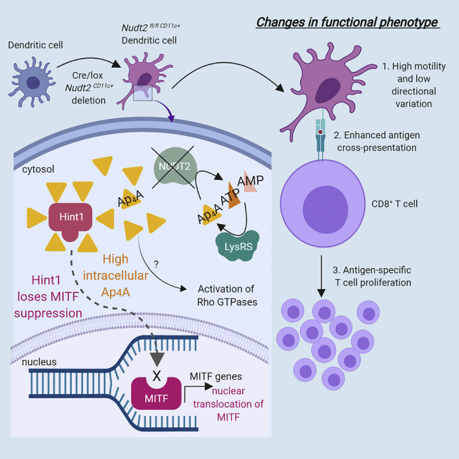
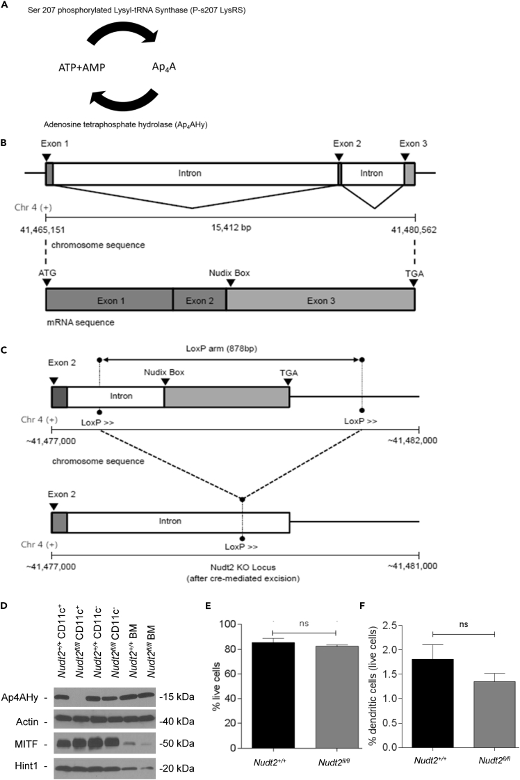
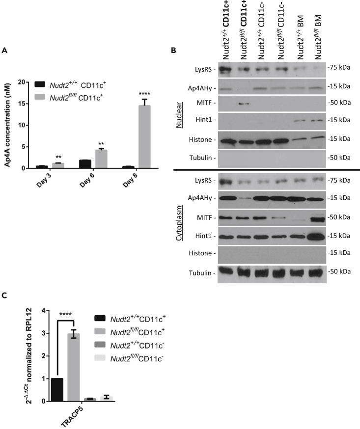
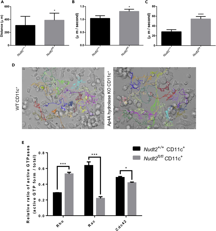
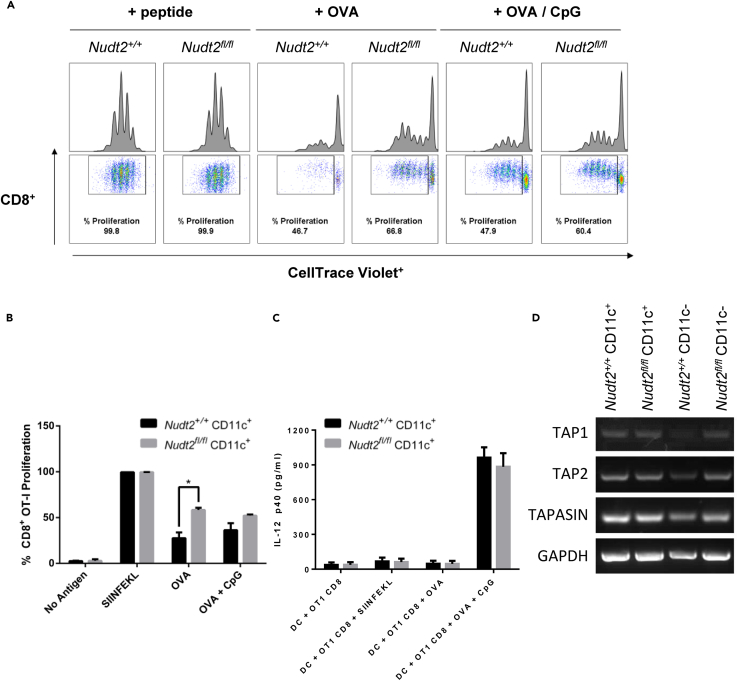
The significance of intracellular Ap4A levels over immune activity of dendritic cells (DCs) has been studied in Nudt2fl/fl/CD11c-cre mice. The transgenic mice have been generated by crossing floxed NUDT2 gene mice with DC marker CD11c recombinase (cre) mice. The DCs derived from these mice have higher levels of Ap4A (≈30-fold) compared with those derived from Nudt2+/+ mice. Interestingly, the elevated Ap4A in DCs has led them to possess higher motility and lower directional variability. In addition, the DCs are able to enhance immune protection indicated by the higher cross-presentation of antigen and priming of CD8+ OT-I T cells. Overall, the study denotes prominent impact of Ap4A over the functionality of DCs. The Nudt2fl/fl/CD11c-cre mice could serve as a useful tool to study the influence of Ap4A in the critical immune mechanisms of DCs.

Keywords: Immune Response; Immunology; Model Organism; Molecular Mechanism of Behavior.


Figures

None
Graphical abstract
Figure 1
Figure 1
Generation of Ap4A Hydrolase Knockout Mice and Viability of Dendritic Cells (A) The phosphorylated form of Lysyl-tRNA transferase (phosphoSerine 207 LysRS) synthesizes Ap4A from ATP and AMP, and is in turn broken down into same by Ap4A hydrolase (Ap4AHy). (B) The gene locus and mRNA structure of NUDT2. The full open reading frame for Ap4AHy (15,412 bp) consists of three exons interspersed with two introns. The Nudix box is nothing but the enzymatically functional site for Ap4AHy and is located at exon 3. (C) The deletion of Ap4AHy by gene floxing and deletion strategy. The two loxP sites were generated on either side of the gene encompassing the full sequence of exon 3 and a portion of immediate intron upstream to it. (D–F) (D) Western blot showing expression levels of Ap4AHy, MITF, and Hint1 in splenic dendritic cells of Nudt2+/+ and Nudt2fl/fl/CD11c+ (Ap4A hydrolase knockout) mice (represents four independent experiments). Immature bone marrow-derived cells (BM). (E) and (F) Viability percentage of isolated BMDCs and splenic DCs. Results (mean ± SEM) represent two independent experiments. The significant difference of test in comparision to control. nsp> 0.05 (Student's t test).
Figure 2
Figure 2
Detailed Functional Characterization of Nudt2fl/fl/CD11c-cre DC (A) Intracellular concentrations of Ap4A in Nudt2+/+ and Nudt2fl/fl/CD11c-cre BMDC were generated from bone marrow cultured with granulocyte-macrophage colony-stimulating factor and examined on days 3, 6, and 8, using a luciferase assay. (B and C) (B) Western blot of LysRS, Ap4AHy, MITF, and Hint1 in Nudt2+/+ and Nudt2fl/fl/CD11c-cre CD11c+ splenic cells. Immature bone marrow-derived cells (BM) (C). Real-time PCR result of MITF-specific gene (TRACP5) expression in BMDCs. The results (mean ± SEM) are representative of four independent experiments and the significant difference of test in comparison to control. **p < 0.01; ****p < 0.0001 (Mann-Whitney test for multiple comparison).
Figure 3
Figure 3
Comparative Motility of DCs from Nudt2fl/fl/CD11c+ and Nudt2+/+ Mice (A) Live imaging microscopy of granulocyte-macrophage colony-stimulating factor-treated DCs (8 days). The post-treated DCs on observational Petri dish were chosen randomly (n = 20) and traced minute wise (time lap ≈30 min) to find the change in cell movement and the distance moved. (B) The speed of cellular movement and the ratio between the distance moved and difference of two time frames. (C) Cellular displacement. (D) A still frame taken from the video created for live cell imaging of Nudt2+/+ and Nudt2fl/fl/CD11c+ DCs. (E) Small GTPase activation state in Nudt2+/+ and Nudt2fl/fl/CD11c+ DCs (n = 2). Results (mean ± SEM) represent two independent experiments. The significant difference of test in comparision to control. *p < 0.05, ***p < 0.001, and ****p < 0.0001 (Student's t test or Mann-Whitney test for multiple comparison).
Figure 4
Figure 4
Nudt2fl/fl/CD11c+ DCs' Potential in Influencing OT-I CD8+ T Cells Antigen Cross-Priming (A) The percentage of OT-I CD8+ T cells that have proliferated following co-culture for 3 days either with Nudt2fl/fl/CD11c+ BMDCs or Nudt2+/+ CD11c+ BMDCs (1:10) incubated with SIINFEKL peptide, OVA and CpG ODN. The harvested cells were analyzed by flow cytometry to quantify CD8+ CellTrace Violet+ population for proliferation. (B) Graph represents the percentage of OT-I CD8+ T cells proliferated when cultured with Nudt2+/+ or Nudt2fl/fl/CD11c+ DCs in response to different antigenic preparations. (C) IL-12 p40 production by DCs during the above co-culture conditions. (D) mRNA levels for TAP1, TAP2, and TAPASIN in Nudt2+/+ and Nudt2fl/fl/CD11c-cre in splenic DCs measured by RT-PCR. Results (mean ± SEM) represent three independent experiments and the significant difference of test in comparision to control. *p < 0.05 (Student's t test or Mann-Whitney test for multiple comparison).

References

    1. Alvarez D., Vollmann E.H., von Andrian U.H. Mechanisms and consequences of dendritic cell migration. Immunity. 2008;29:325–342. - PMC - PubMed
    2. Alvarez, D., Vollmann, E.H., and von Andrian, U.H.. (2008). Mechanisms and consequences of dendritic cell migration. Immunity 29, 325-342. - PMC - PubMed
    1. Carmi-Levy I., Yannay-Cohen N., Kay G., Razin E., Nechushtan H. Diadenosine tetraphosphate hydrolase is part of the transcriptional regulation network in immunologically activated mast cells. Mol. Cell. Biol. 2008;28:5777–5784. - PMC - PubMed
    2. Carmi-Levy, I., Yannay-Cohen, N., Kay, G., Razin, E., and Nechushtan, H.. (2008). Diadenosine tetraphosphate hydrolase is part of the transcriptional regulation network in immunologically activated mast cells. Mol. Cell. Biol. 28, 5777-5784. - PMC - PubMed
    1. Carmi-Levy I., Motzik A., Ofir-Birin Y., Yagil Z., Yang C.M., Kemeny D.M., Han J.M., Kim S., Kay G., Nechushtan H. Importin beta plays an essential role in the regulation of the LysRS-Ap(4)A pathway in immunologically activated mast cells. Mol. Cell. Biol. 2011;31:2111–2121. - PMC - PubMed
    2. Carmi-Levy, I., Motzik, A., Ofir-Birin, Y., Yagil, Z., Yang, C.M., Kemeny, D.M., Han, J.M., Kim, S., Kay, G., Nechushtan, H., et al. (2011). Importin beta plays an essential role in the regulation of the LysRS-Ap(4)A pathway in immunologically activated mast cells. Mol. Cell. Biol. 31, 2111-2121. - PMC - PubMed
    1. Carracedo G., Guzman-Aranguez A., Loma P., Pintor J. Diadenosine polyphosphates release by human corneal epithelium. Exp. Eye Res. 2013;113:156–161. - PubMed
    2. Carracedo, G., Guzman-Aranguez, A., Loma, P., and Pintor, J.. (2013). Diadenosine polyphosphates release by human corneal epithelium. Exp. Eye Res. 113, 156-161. - PubMed
    1. Castany M., Jordi I., Catala J., Gual A., Morales M., Gasull X., Pintor J. Glaucoma patients present increased levels of diadenosine tetraphosphate, Ap4A, in the aqueous humour. Exp. Eye Res. 2011;92:221–226. - PubMed
    2. Castany, M., Jordi, I., Catala, J., Gual, A., Morales, M., Gasull, X., and Pintor, J. (2011). Glaucoma patients present increased levels of diadenosine tetraphosphate, Ap4A, in the aqueous humour. Exp. Eye Res. 92, 221-226. - PubMed