. 2019 Sep:105:35-45.
doi: 10.1016/j.archoralbio.2019.05.015. Epub 2019 May 20.

The influence of masseter and temporalis sarcomere length operating ranges as determined by laser diffraction on architectural estimates of muscle force and excursion in macaques (Macaca fascicularis and Macaca mulatta)

Affiliations

The influence of masseter and temporalis sarcomere length operating ranges as determined by laser diffraction on architectural estimates of muscle force and excursion in macaques (Macaca fascicularis and Macaca mulatta)

Andrea B Taylor et al. Arch Oral Biol. 2019 Sep.

Abstract

Objective: Determine sarcomere length (Ls) operating ranges of the superficial masseter and temporalis in vitro in a macaque model and examine the impact of position-dependent variation on Ls and architectural estimates of muscle function (i.e., fiber length, PCSA) before and after Ls-normalization.

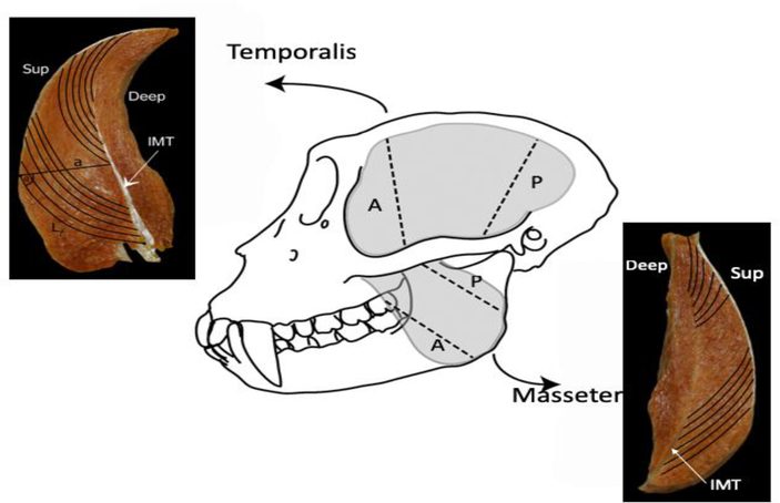
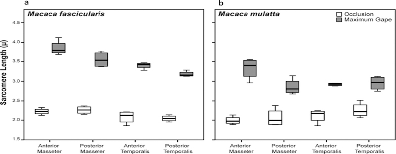
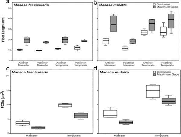
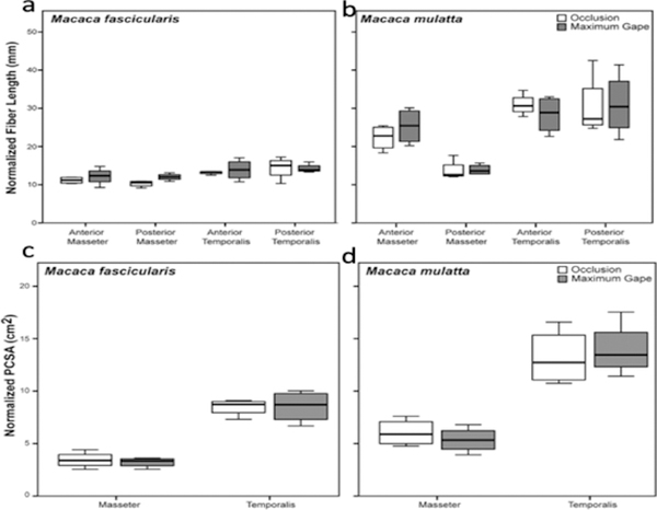
Design: Heads of adult Macaca fascicularis (n = 4) and M. mulatta (n = 3) were bisected postmortem. One side of the jaw was fixed in occlusion, the other in maximum gape. Ls was measured bilaterally using laser diffraction and these measurements were used to estimate sarcomere-length operating ranges. Differences in fiber length and PCSA between sides were tested for significance prior to and following Ls-normalization.

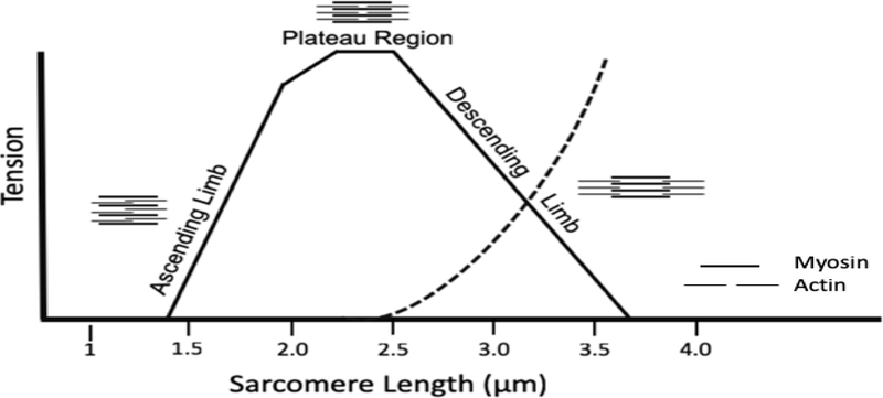
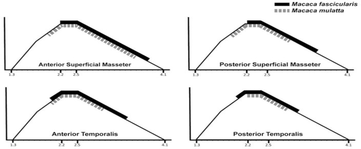
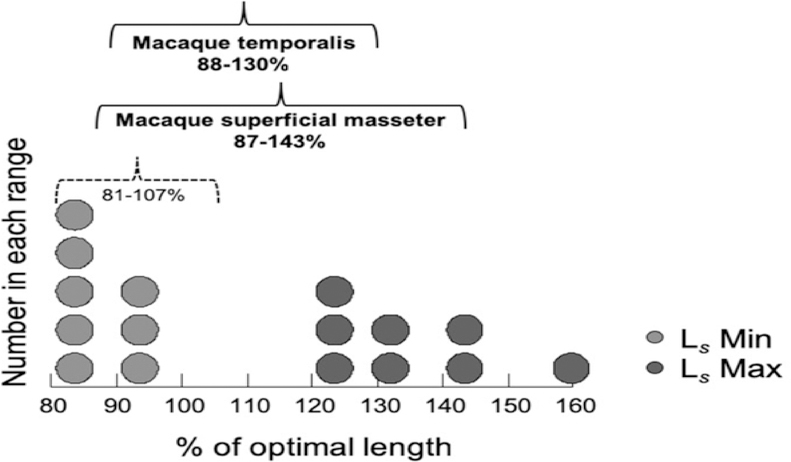
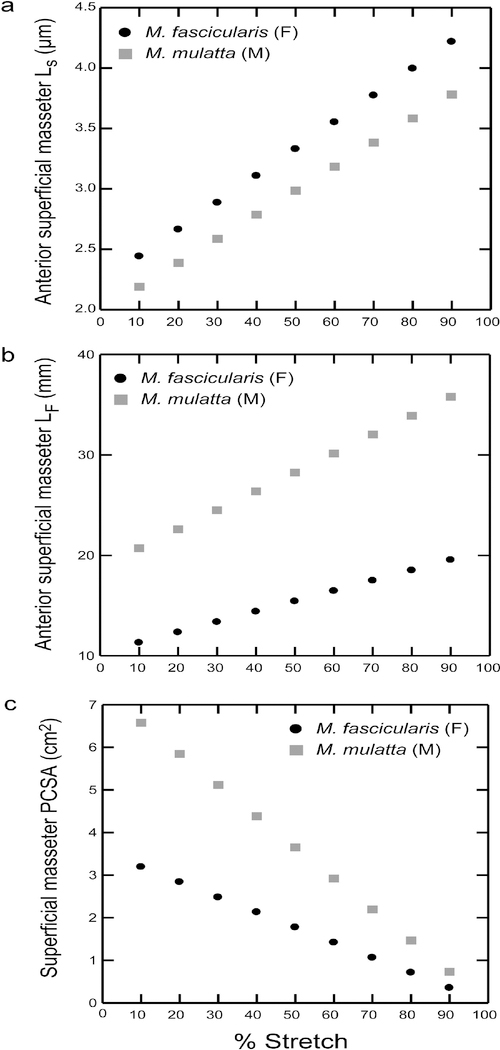
Results: Sarcomere-length operating ranges were widest for the anterior superficial masseter and narrowest for the posterior temporalis. Compared with other mammals, macaque operating ranges were wider and shifted to the right of the descending limb of a representative length-tension curve. Fibers were significantly stretched by as much as 100%, and PCSAs reduced by as much as 43%, on the maximally gaped compared with occluded sides. Ls-normalization substantially reduced position-dependent variance.

Conclusions: The superficial masseter ranges between 87-143% and the temporalis between 88-130% of optimal Ls from maximum gape to occlusion, indicating maximum relative Ls for these macaque muscles exceeds the upper end range previously reported for the jaw muscles of smaller mammals. The wider macaque operating ranges may be functionally linked to the propensity for facially prognathic primates to engage in agonistic canine display behaviors that require jaw-muscle stretch to facilitate production of wide jaw gapes.

Keywords: Fiber length; Macaque; Masseter; Sarcomere length; Temporalis.


Conflict of interest statement

Conflict of interest

None declared.

Figures

Figure 1.
Figure 1.
Schematic of a representative sarcomere length-tension curve for skeletal muscle. Myosin (thick) and actin (thin) filaments show varying amounts of overlap in different regions of the length-tension curve. Tension diminishes as myofilament overlap increases (Ascending Limb) or decreases (Descending Limb) relative to the Plateau Region. Dotted line represents passive tension.
Figure 2.
Figure 2.
Schematic of a macaque skull (M. fascicularis). The gray-shaded areas represent the temporalis and masseter muscles in situ. Once removed en masse from the skull, each muscle was sectioned along its length into anterior and posterior regions (black hatched lines) for fiber length measurements. Cross sections of each muscle depict measurements taken at each section: Lf, fiber length; a, perpendicular distance from the intramuscular tendon [IMT] to the superficial tendon. Pinnation angle [θ] was computed as the arcsine of a/NLf.
Figure 3.
Figure 3.
Box plots of masseter and temporalis sarcomere length measurements taken at occlusion and maximum jaw gape for a) M. fascicularis and b) M. mulatta. Sarcomere lengths at maximum jaw gape are significantly (P < 0.05) greater compared with sarcomere lengths at occlusion in both species for all muscle regions (see Table 2). Sarcomere-length operating range is greatest for the anterior superficial masseter in both species. In this and all subsequent box plots, line within box denotes median. Boundaries of the box represent 25th and 75th percentiles. Whiskers indicate 10th and 90th percentiles.
Figure 4.
Figure 4.
Masseter and temporalis sarcomere-length operating ranges for M. fascicularis (solid black line) and M. mulatta (hatched grey line) superimposed on a length-tension curve for rhesus macaque myofilament lengths (Walker and Schrodt, 1974). Operating range is interpolated from Ls values at occlusion and maximum gape. Male Macaca mulatta sarcomere lengths operate over narrower ranges and are shifted to the left compared with female M. fascicularis, reflecting the longer male fibers. With more sarcomeres in series to minimize the amount of stretch during angular rotation of the joint, fibers remain closer to the peak of the length-tension curve, minimizing loss of force.
Figure 5.
Figure 5.
Box plots of masseter and temporalis fiber lengths (a-b) and PCSAs (c-d) at occlusion and maximum jaw gape. Fibers are significantly longer (P < 0.05) and PCSAs significantly smaller (P < 0.05) at maximum jaw gape compared with fibers and PCSAs at occlusion in both species for all muscle regions (see Table 3). As fibers are stretched (and pinnation angles decrease; Supplementary Table S2), muscle PCSAs decrease as a function of change in jaw position.
Figure 6.
Figure 6.
Box plots of masseter and temporalis fiber lengths (a-b) and PCSAs (c-d) at occlusion and maximum jaw gape, normalized to a standard sarcomere length of 2.41 μm. The absence of significant differences (P > 0.05) in fiber lengths and PCSAs between the occluded and maximally-gaped sides indicates that Ls-normalization has effectively eliminated the impact of position-dependent variation in fiber lengths and PCSAs in these jaw muscles (see Table 4).
Figure 7.
Figure 7.
Dot density histogram of relative sarcomere lengths (measured sarcomere length divided by a standard sarcomere length of 2.41 μm; reported as % of optimal length, L0) for macaque anterior and posterior superficial masseter and anterior and posterior temporalis muscles. Light grey dots represent relative Ls Min (at occlusion) and dark grey dots represent relative Ls Max (at maximum jaw gape), for each muscle region, averaged by species. The average range is 87–143% L0 for macaque masseter and 88–130% L0 for the temporalis (solid black brackets). The range for relative sarcomere lengths for these macaque jaw-adductors includes a large portion of the descending limb of the L-T curve, and is both wider and shifted to the right of the L-T curve in comparison with the range reported for jaw and limb muscles (81–107%; dotted black bracket) across a variety of vertebrates (Burkholder & Lieber, 2001). The macaque pattern may be characteristic of facially prognathic primates and other mammals that generate relatively wide maximum jaw gapes.
Figure 8.
Figure 8.
Plots of changing a) sarcomere lengths (Ls); b) fiber length (Lf); and c) PCSA as a function of percent fiber stretch for the anterior superficial masseter. Fiber stretch is interpolated in 10% increments from occlusion to 90%. For every 10% increase in stretch from occlusion, sarcomeres lengthen by ~8–9%, and fibers lengthen between 1–3 mm, depending on muscle region. Sarcomere and fiber lengths measured in vitro at maximum jaw gapes fall within our estimated ranges for sarcomeres stretched by as much as 70–80%, and fiber stretched by as much as 90%, respectively, from occlusion. (Supplementary Table S3).

References

    1. Anapol F, & Barry K (1996). Fiber architecture of the extensors of the hindlimb in semiterrestrial and arboreal guenons. American Journal of Physical Anthropology, 99, 429–447. - PubMed
    1. Anapol F, & Gray JP (2003). Fiber architecture of the intrinsic muscles of the shoulder and arm in semiterrestrial and arboreal guenons. American Journal of Physical Anthropology, 122, 51–65. - PubMed
    1. Anapol F, & Jungers WL (1986). Architectural and histochemical diversity within the quadriceps femoris of the brown lemur (Lemur fulvus). American Journal of Physical Anthropology, 69, 355–375. - PubMed
    1. Anapol F, Shahnoor N, & Gray JP (2004). Fiber architecture, muscle function, and behavior: Gluteal and hamstring muscles of semiterrestrial and arboreal guenons In Anapol F, German RZ & Jablonski NG (Eds.), Shaping Primate Evolution (pp. 99–133). Cambridge: Cambridge Studies in Biological and Evolutionary Anthropology.
    1. Anapol F, Shahnoor N, & Ross CF (2008). Scaling of reduced physiologic cross-sectional area in primate muscles of mastication In Vinyard CJ, Ravosa MJ & Wall CE (Eds.), Primate Craniofacial Function and Biology (pp. 201–215). New York: Springer.