. 2019 Aug 21;103(4):627-641.e7.
doi: 10.1016/j.neuron.2019.05.035. Epub 2019 Jun 26.

Transneuronal Propagation of Pathologic α-Synuclein from the Gut to the Brain Models Parkinson's Disease

Affiliations

Transneuronal Propagation of Pathologic α-Synuclein from the Gut to the Brain Models Parkinson's Disease

Sangjune Kim et al. Neuron. .

Abstract

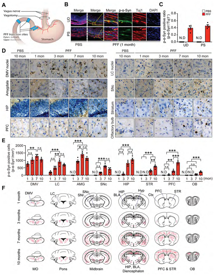
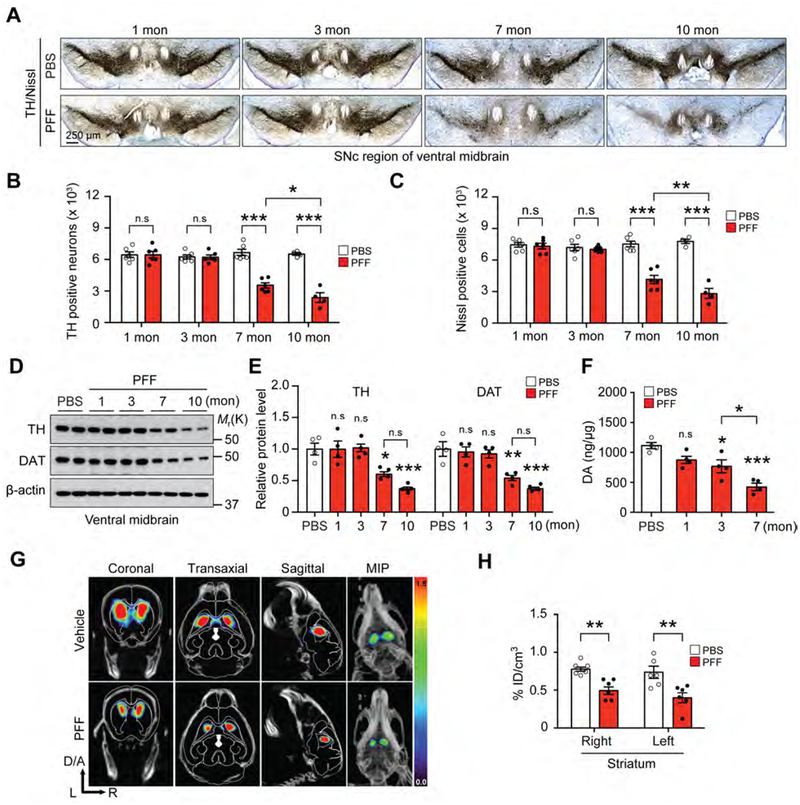
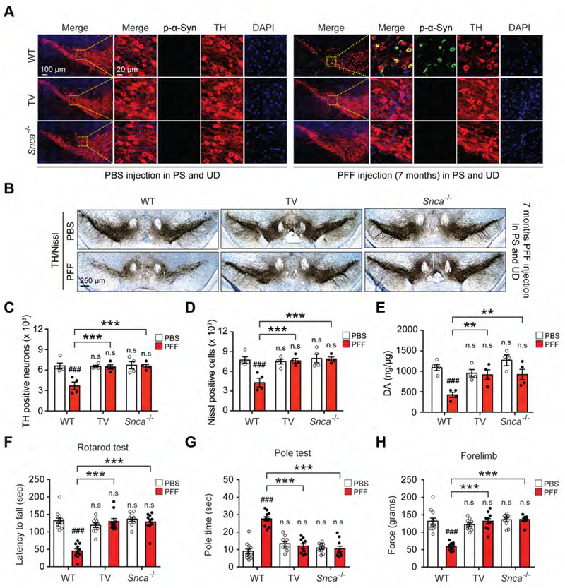
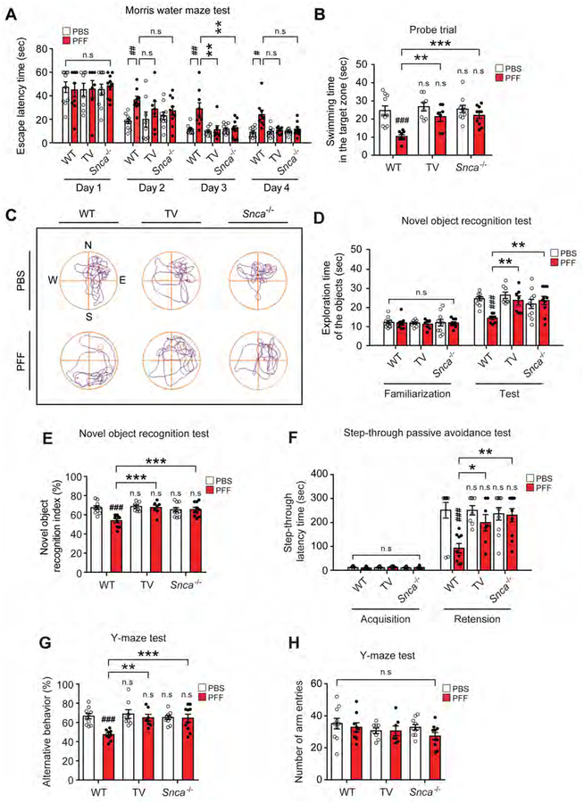
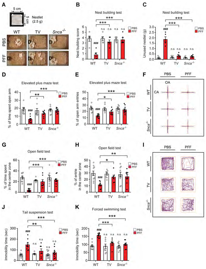
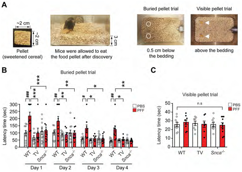
Analysis of human pathology led Braak to postulate that α-synuclein (α-syn) pathology could spread from the gut to brain via the vagus nerve. Here, we test this postulate by assessing α-synucleinopathy in the brain in a novel gut-to-brain α-syn transmission mouse model, where pathological α-syn preformed fibrils were injected into the duodenal and pyloric muscularis layer. Spread of pathologic α-syn in brain, as assessed by phosphorylation of serine 129 of α-syn, was observed first in the dorsal motor nucleus, then in caudal portions of the hindbrain, including the locus coeruleus, and much later in basolateral amygdala, dorsal raphe nucleus, and the substantia nigra pars compacta. Moreover, loss of dopaminergic neurons and motor and non-motor symptoms were observed in a similar temporal manner. Truncal vagotomy and α-syn deficiency prevented the gut-to-brain spread of α-synucleinopathy and associated neurodegeneration and behavioral deficits. This study supports the Braak hypothesis in the etiology of idiopathic Parkinson's disease (PD).

Keywords: Braak hypothesis; Lewy body pathology; Parkinson’s disease; gut to brain transmission; motor symptoms; neurodegeneration; non-motor symptoms; pre-formed fibrils; vagus nerve; α-synuclein.


Conflict of interest statement

DECLARATION OF INTERESTS

The authors declare no competing interests.

Figures

Figure 1.
Figure 1.. α-syn PFF injection into the gut triggers progressive spreading of α-syn pathology to the enteric nervous system followed by spread to connected brain regions.
(A) Overview of the site of injection and vagotomy (B) Representative double-immunostaining for pSer129-α-syn (white) and Tuj-1 (red) in the upper duodenum (UD) and pyloric stomach (PS) after 1 month post-injection. (C) Quantification of pSer129-α-syn positive neurons normalized to Tuj-1 positive neurons in the upper duodenum and pyloric stomach (n=4). (D) Brain distribution of pSer129-α-syn accumulation in mice that received α-syn PFF in the gut. pSer129-α-syn immunohistochemistry from the dorsal motor nucleus of the vagus to the olfactory bulb of α-syn PFF gastrointestinal injected mice sacrificed at 1, 3, 7, and 10 months post-injection. (E) Quantification of pSer129-α-syn immunoreactivity shown in panel D (n=4). (F) Diagram illustrating the CNS distribution of pSer129-α-syn representing α-syn pathology (red dots) in the brain from coronal sections from 1, 3, 7, and 10 months post-injection. Error bars represent the mean ± S.E.M. Statistical significance was determined using a two-way ANOVA followed by post-hoc Bonferroni test for multiple group comparison. ***P < 0.001. N.D: not detected. BLA, basolateral amygdala; CPu, upper caudate-putamen; Ctx, cortex; DG, dentate gyrus; DMV, dorsal motor nucleus of the vagus; HIP, hippocampus; LC, locus coeruleus; MO, medulla oblongata; OB, olfactory bulb; PFC, prefrontal cortex; PS, pyloric stomach; SNc. substantia nigra pars compacta; SNr, substantia nigra pars reticulate; STR, striatum; UD, upper duodenum. See also Figures S1, S2 and S3.
Figure 2.
Figure 2.. α-syn PFF injection into the gut leads to progressive PD-like pathology.
(A) Representative photomicrographs from coronal mesencephalon sections containing TH-positive neurons in the SNc region of 1, 3, 7, and 10 months after gastrointestinal injection of α-syn PFF. (B) Stereology counts of TH and (C) Nissl-positive neurons in the SNc region of one hemisphere. Unbiased stereological counting was performed in the SNc region (n=4–6). (D) Representative immunoblots of TH, DAT, and β-actin in the ventral midbrain over time. (E) Quantification of TH, and DAT protein levels normalized to β-actin (n=4). (F) DA concentrations in the STR of PBS and α-syn PFF gastrointestinal injected mice over time as measured by HPLC (n=4). (G) DAT SPECT/CT scans showing representative images after PBS and α-syn PFF gastrointestinal injected mice at 7 months. (H) Quantification of DAT SPECT/CT results (n=6). Error bars represent the mean ± S.E.M. Statistical significance was determined using a two-way ANOVA followed by post-hoc Bonferroni test for multiple group comparison. *P< 0.05, **P < 0.01, ***P < 0.001. n.s: not significant. See also Figure S3.
Figure 3.
Figure 3.. Vagotomy and α-syn deficiency prevents PD-like pathology induced by α-syn PFF injection into the gut.
(A) Representative double-immunostaining for pSer129-α-syn (white) and TH (red) in SNc. (B) Representative photomicrographs from coronal mesencephalon sections containing TH-positive neurons in the SNc region of 7 months after α-syn PFF gastrointestinal injected WT, vagotomy (TV) and Snca−/− mice. (C) Stereology counts of TH and (D) Nissl-positive neurons in the SNc region of one hemisphere. Unbiased stereological counting was performed in the SNc region of WT, TV, and Snca−/− mice (n=4). (E) DA concentrations in the STR of PBS and α-syn PFF gastrointestinal injected WT, vagotomy and Snca−/− mice as determined by HPLC (n=4). (F-H) Behavioral assessment at 7 months after PBS and α-syn PFF gastrointestinal injected WT (n=11–12), TV (n=10) and Snca−/− mice (n=10). Results of mice on the (F) rotarod test, (G) pole test, and (H) forelimb grip strength test. Error bars represent the mean ± S.E.M. Statistical significance was determined using a two-way ANOVA followed by post-hoc Bonferroni test for multiple group comparison. ###P < 0.001 vs. PBS gastrointestinal injected WT group. **P < 0.01, ***P < 0.001 vs. α-syn PFF gastrointestinal injected WT mice group. n.s: not significant. See also Figures S4 and S5 and Movie S1.
Figure 4.
Figure 4.. Vagotomy and α-syn deficiency prevents cognitive deficits induced by α-syn PFF injection into the gut.
Cognitive behavioral assessments 7 months after PBS and α-syn PFF gastrointestinal injection in WT (n=9–10), vagotomy (TV) (n=7–8) and Snca−/− mice (n=10). (A) Escape latency time and (B) probe trial session in the Morris water maze test. (C) Representative swimming paths of mice from each group in the MWMT on the probe trial day 5. The mice were then given two trial sessions each day for four consecutive days, with an inter-trial interval of 15 min, and the escape latencies were recorded. This parameter was averaged for each session of trials and for each mouse. (D) Exploration time of the objects and (E) percentage of novel object recognition index in the novel object recognition test. (F) Effect of TV and α-syn deficiency on α-syn PFF-induced hippocampal-dependent contextual or amygdala-dependent fear conditioning learning and memory in the step-through passive avoidance test. The time taken for a mouse to enter the dark compartment after door opening was defined as latency for both training and test trials. (G-H) Effect of TV and α-syn deficiency on α-syn PFF-induced short-term or working memory in the Y-maze test. (G) Percentage of alternative behavior and (H) number of arm entries in the Y-maze test. Error bars represent the mean ± S.E.M. All behavior tests were analyzed by two-way ANOVA followed by post-hoc Bonferroni test for multiple group comparison. ##P < 0.01, ###P < 0.001 vs. PBS gastrointestinal injected WT group. *P< 0.05, **P < 0.01, ***P < 0.001 vs. α-syn PFF gastrointestinal injected WT group. n.s: not significant. See also Figure S6 and Movies S2.
Figure 5.
Figure 5.. Vagotomy and α-syn deficiency prevent deficits in psychological behavior induced by α-syn PFF injection into the gut.
Behavioral assessments of psychological behavior at 7 months after PBS and α-syn PFF gastrointestinal injection were performed in WT (n=9–10), TV (n=7–8) and Snca−/− mice (n=10). (A) Representative images of nest building. Images show nest building after 16 h following introduction of nestlet in among all experimental groups. (B) Nest building score and (C) unused nestlet in the nest building test. Nest-building scores were assessed, and the amount of unused nestlet material was measured after 16 h. (D) Percentage of time spent in the open arm and (E) Open arm entries in the EPM. (F) Representative movement paths of mice from each group in the EPM. (G-I) Effect of TV and α-syn deficiency on α-syn PFF-induced locomotion and central activity in the OFT. The data of percentage of (G) time spent and (H) entries in the center zone in the OFT. (I) Representative movement paths of mice from each group in the OFT. Effect of vagotomy and α-syn deficiency on α-syn PFF-induced depressive-like behavior in the (J) TST and (K) FST. The immobility times were recorded using the video tracking system (ANY-Maze software) during final 4-min of a total 6 min test. Error bars represent the mean ± S.E.M. All behavior tests were analyzed by two-way ANOVA followed by post-hoc Bonferroni test for multiple group comparison. ###P < 0.001 vs. PBS gastrointestinal injected WT group. *P< 0.05, **P < 0.01, ***P < 0.001 vs. α-syn PFF gastrointestinal injected WT group. n.s: not significant. See also Figure S6, Movies S3, S4 and S5.
Figure 6.
Figure 6.. Vagotomy and α-syn deficiency prevent olfactory dysfunctions induced by α-syn PFF injection into the gut.
(A-C) Olfactory behavioral assessments at 9 months after PBS and α-syn PFF gastrointestinal injection in WT (n=9–10), TV (n=7–8) and Snca−/− mice (n=10). The buried pellet trial test was performed for 4 days and the surface pellet control test was performed for one trial 1 day after the buried pellets. (A) Two pieces of sweetened cereal were buried along the perimeter of the cage approximately 0.5 cm below the bedding so that it was not visible. (B) Latency in finding the first pellet was recorded when the mouse touched the pellet. (C) The visible pellet trial test was set up in a similar way except that the piece of cereal was placed on top of the bedding. Error bars represent the mean ± S.E.M. Statistical significance was determined using a two-way ANOVA followed by post-hoc Bonferroni test for multiple group comparison. #P < 0.05, ##P < 0.01, ###P < 0.001 vs. PBS gastrointestinal injected WT group. *P< 0.05, **P < 0.01, ***P < 0.001 vs. α-syn PFF gastrointestinal injected WT group. n.s: not significant.

Comment in

References

    1. Anderson JP, Walker DE, Goldstein JM, de Laat R, Banducci K, Caccavello RJ, Barbour R, Huang J, Kling K, Lee M, et al. (2006). Phosphorylation of Ser-129 is the dominant pathological modification of alpha-synuclein in familial and sporadic Lewy body disease. J Biol Chem 281, 29739–29752. - PubMed
    1. Arias N, Mendez M, and Arias JL (2015). The importance of the context in the hippocampus and brain related areas throughout the performance of a fear conditioning task. Hippocampus 25, 1242–1249. - PubMed
    1. Bernhardt N, Lieser MK, Hlusicka EB, Habelt B, Wieske F, Edemann-Callesen H, Garthe A, and Winter C (2018). Learning deficits in rats overexpressing the dopamine transporter. Sci Rep 8, 14173. - PMC - PubMed
    1. Berthoud HR, Carlson NR, and Powley TL (1991). Topography of efferent vagal innervation of the rat gastrointestinal tract. Am J Physiol 260, R200–207. - PubMed
    1. Bevins RA, and Besheer J (2006). Object recognition in rats and mice: a one-trial-non-matching-to-sample learning task to study ‘recognition memory’. Nat Protoc 1, 1306–1311. - PubMed

Publication types

MeSH terms