. 2019 Jun 30:25:4837-4848.
doi: 10.12659/MSM.916311.

Helicobacter Pylori Induces GATA3-Dependent Chitinase 3 Like 1 (CHI3L1) Upregulation and Contributes to Vascular Endothelial Injuries

Affiliations

Helicobacter Pylori Induces GATA3-Dependent Chitinase 3 Like 1 (CHI3L1) Upregulation and Contributes to Vascular Endothelial Injuries

Jingshu Chi et al. Med Sci Monit. .

Abstract

BACKGROUND Helicobacter pylori infection is associated with various vascular diseases. However, its mechanism is yet to be defined. The present study aimed to investigate the effect of H. pylori on vascular endothelial cells as well as the GATA3-related mechanism of H. pylori infection-induced endothelial injuries. MATERIAL AND METHODS A co-culture of H. pylori with human umbilical endothelial cells (HUVECs) was produced. The proliferation of HUVECs that had been incubated with H. pylori were examined via CCK-8 (Cell Counting Kit-8) and EdU (5-ethynyl-2'-deoxyuridine) staining. Cell migration and microtubules formation were studied using Transwell and tube formation respectively. Construction of a mouse model of H. pylori infection as well as the expression of GATA3 and CHI3L1 in vessels were tested using western blot and immunohistochemistry. Small interfering RNA (siRNA) of GATA3 were transfected into HUVECs in order to establish cell lines with knocked-down GATA3. The production of the aforementioned molecules and p38 mitogen-activated protein kinase (MAPK) related molecules in HUVECs was measured using quantitative real-time polymerase chain reaction and western blot. RESULTS H. pylori significantly inhibited the proliferation, migration, and tube formation of HUVECs, as well as increased the production of the inflammatory factor CHI3L1 and phosphorylated p38 from endothelial cells along with an increased expression of GATA3. Elevated levels of the GATA3 and CHI3L1 were also found in the arteries of H. pylori-infected mice. Following GATA3 knockdown, the H. pylori-induced dysfunction of HUVECs was restored. CONCLUSIONS H. pylori impaired vascular endothelial function. This might be due to the H. pylori-induced increased expression of GATA3, as well as the GATA3 mediated upregulated CHI3L1 and activation of the p38 MAPK pathway.


Conflict of interest statement

Conflicts of interest

None.

Figures

Figure 1
Figure 1
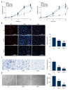
Helicobacter pylori inhibited the biological functions of HUVECs. (A) CCK-8 assay was used to evaluate the proliferation of the HUVECs after being co-cultured with the CagA H. pylori or CagA+ H. pylori at the MOI=50, 100, 150, or no H. pylori. (B) EdU experiments were used to further verify the proliferation of HUVECs inhibited after being co-cultured with the CagA H. pylori or CagA+ H. pylori at MOI=150 for 48 hours. (C) Transwell experiments demonstrated the migration function of HUVECs was declined after being co-cultured with H. pylori. (D) Tube formation assays showed the ability of forming tubular structures of HUVECs was decreased after being co-cultured with H. pylori. ** P<0.01, *** P<0.001, **** P<0.0001 versus NC group. # P<0.05, #### P<0.0001 versus CagA group. HUVECs, human umbilical endothelial cells; CCK-8 – Cell Counting Kit-8; CagA – cytotoxin-related protein; MOI – multiplicity of infection; EdU – 5-ethynyl-2′-deoxyuridine.
Figure 2
Figure 2
The expression of GATA3 and CHI3L1 were upregulated in HUVECs co-cultured with Helicobacter pylori. (A) qRT-PCR was used to show the expression of GATA3 and CHI3LI mRNA in HUVECs after being cultured with medium added with CagA H. pylori or CagA+ H. pylori or PBS for 48 hours. (B, C) Western blot was used to show the elevated expression of GATA3 and CHI3L1 protein of the aforementioned cells, Calnexin levels were used as a loading control. * P<0.05, ** P<0.01, *** P<0.0001 **** P<0.0001 versus NC group. # P<0.05, ## P<0.01 versus CagA group. HUVECs – human umbilical endothelial cells; CagA – cytotoxin-related protein; qRT-PCR – quantitative real-time polymerase chain reaction; PBS – phosphate-buffered saline.
Figure 3
Figure 3
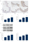
The expression of GATA3 and CHI3L1 were elevated in the Helicobacter pylori infected mice model. (A, B) Immunohistochemical staining was used to demonstrate the upregulation of GATA3 and CHI3L1 in mice thoracic aorta after being gavaged with CagA+ H. pylori or CagA H. pylori. (C, D) Western blot used to show the expression of GATA3 and CHI3L1 protein of the aforementioned mice thoracic aorta, β-asctin levels were used as a loading control. * P<0.05, ** P<0.01, *** P<0.0001 **** P<0.0001 versus NC group. # P<0.05 versus CagA group. CagA – cytotoxin-related protein.
Figure 4
Figure 4
Small interfering RNA (siRNA)-mediated knockdown of GATA3 lowered Helicobacter pylori -induced expression of CHI3L1 and p38 phosphorylation (A) Western blot shows the expression of GATA3 and CHI3L1 protein in HUVECs after transfected with siRNA-control or siRNA-GATA3 for 24 hours and cultured with medium added with CagA H. pylori or CagA+ H. pylori or PBS for 48 hours. Calnexin levels were used as a loading control. (B) The expression of GATA3 was significant reduced by siRNA-GATA3 transfected. (C) The elevated expression of CHI3L1 caused by H. pylori was relatively decreased as GATA3 was knocked down. (D) The H. pylori-induced activation of p38 MAPK was attenuated in GATA3 knocked-down cells. * P<0.05, ** P<0.01, *** P<0.0001 versus si-control group. HUVECs – human umbilical endothelial cells; CagA – cytotoxin-related protein; PBS – phosphate-buffered saline MAPK – mitogen-activated protein kinase.
Figure 5
Figure 5
Knockdown of GATA3 restored the CagA+ Helicobacter pylori-induced inhibitory function in HUVECs. (A) The EdU staining showed the inhibited proliferation by CagA+ H. pylori was restored after GATA3 knockdown. (B) Transwell experiments demonstrated the migration function was increased after GATA3 knockdown. (C) Tube formation ability was partly repaired after GATA3 knockdown. ** P<0.01 versus si-control+CagA+ H group. CagA – cytotoxin-related protein; HUVECs – human umbilical endothelial cells; EdU – 5-ethynyl-2′-deoxyuridine.

References

    1. Lamb A, Chen LF. Role of the Helicobacter pylori-induced inflammatory response in the development of gastric cancer. J Cell Biochem. 2013;114(3):491–97. - PMC - PubMed
    1. Franceschi F, Zuccala G, Roccarina D, Gasbarrini A. Clinical effects of Helicobacter pylori outside the stomach. Nat Rev Gastroenterol Hepatol. 2014;11(4):234–42. - PubMed
    1. Sawayama Y, Ariyama I, Hamada M, et al. Association between chronic Helicobacter pylori infection and acute ischemic stroke: Fukuoka Harasanshin Atherosclerosis Trial (FHAT) Atherosclerosis. 2005;178(2):303–9. - PubMed
    1. Wan Z, Hu L, Hu M, et al. Helicobacter pylori infection and prevalence of high blood pressure among Chinese adults. J Hum Hypertens. 2018;32(2):158–64. - PubMed
    1. Backert S, Tegtmeyer N, Fischer W. Composition, structure and function of the Helicobacter pylori cag pathogenicity island encoded type IV secretion system. Future Microbiol. 2015;10(6):955–65. - PMC - PubMed

MeSH terms