

Role of Mast Cells in Shaping the Tumor Microenvironment
- PMID: 31256327
- PMCID: PMC7244463
- DOI: 10.1007/s12016-019-08753-w
Role of Mast Cells in Shaping the Tumor Microenvironment
Abstract
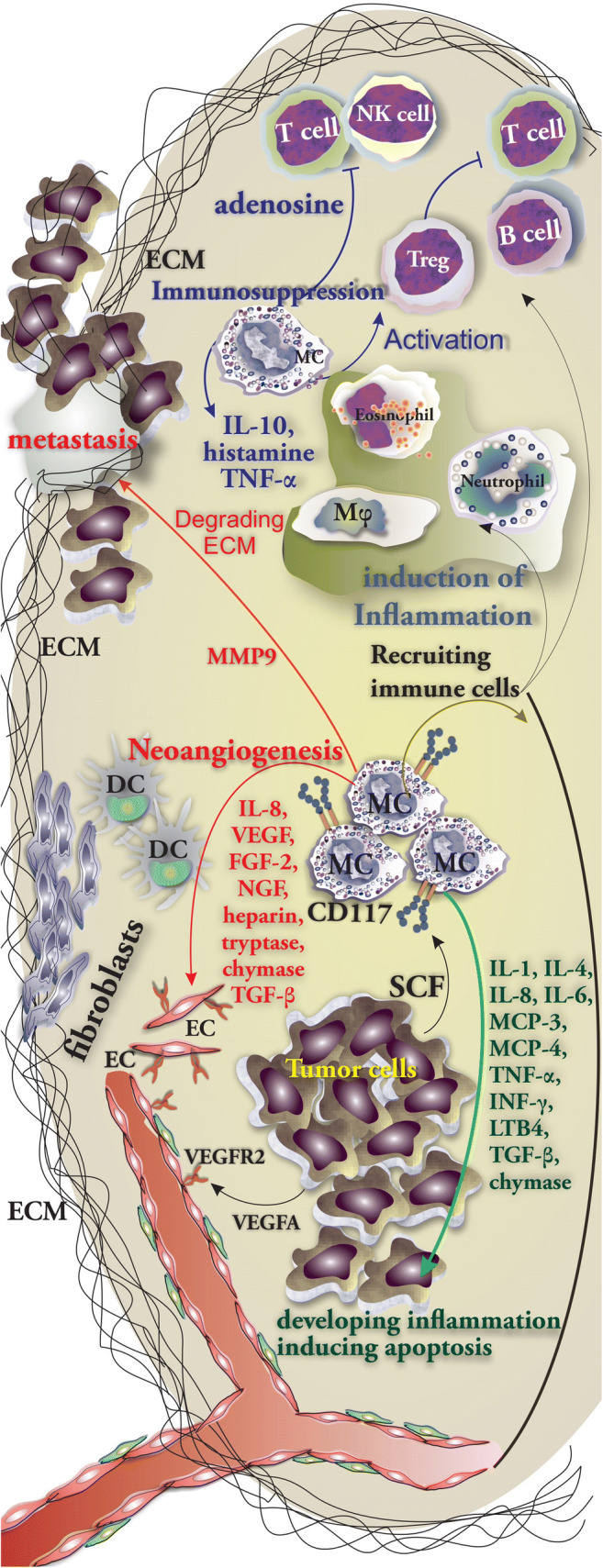
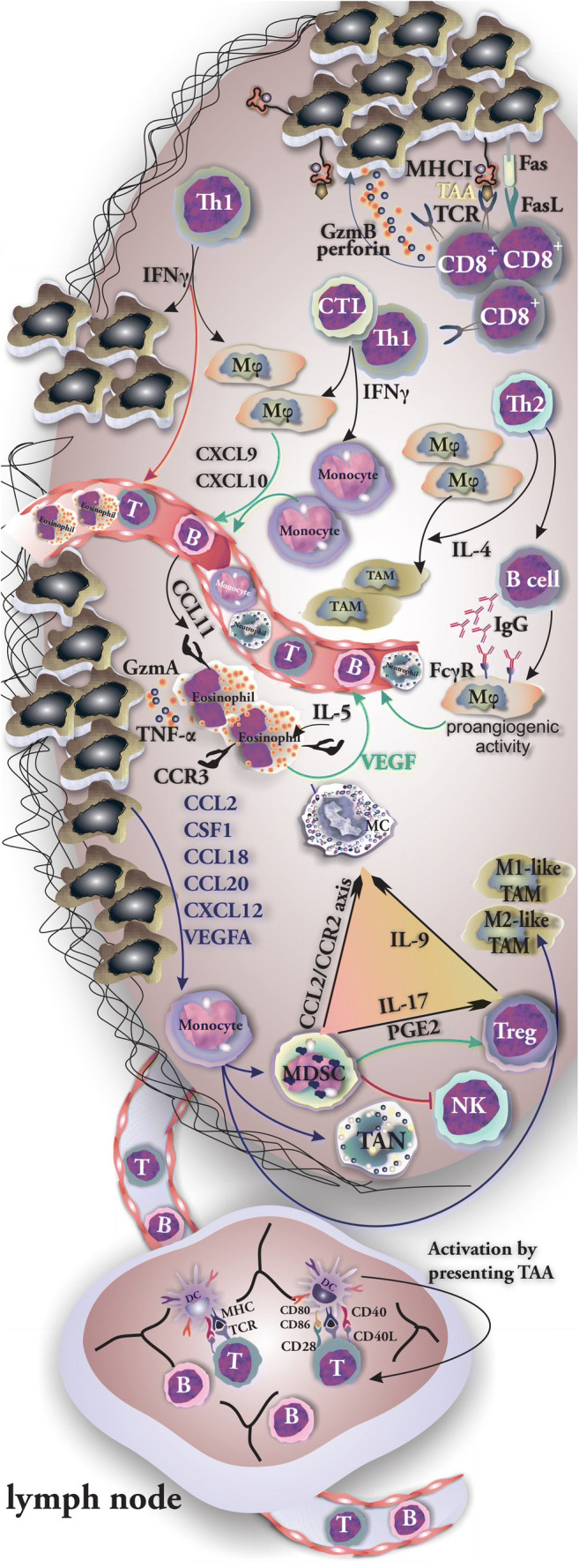
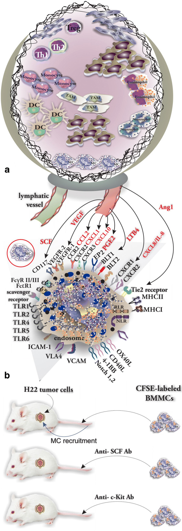
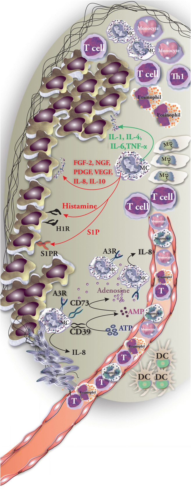
Early mast cell (MC) infiltration has been reported in a wide range of human and animal tumors particularly malignant melanoma and breast and colorectal cancer. The consequences of their presence in the tumor microenvironment (TME) or at their margins still remain unclear as it is associated with a good or poor prognosis based on the type and anatomical site of the tumor. Within the tumor, MC interactions occur with infiltrated immune cells, tumor cells, and extracellular matrix (ECM) through direct cell-to-cell interactions or release of a broad range of mediators capable of remodeling the TME. MCs actively contribute to angiogenesis and induce neovascularization by releasing the classical proangiogenic factors including VEGF, FGF-2, PDGF, and IL-6, and nonclassical proangiogenic factors mainly proteases including tryptase and chymase. MCs support tumor invasiveness by releasing a broad range of matrix metalloproteinases (MMPs). MC presence within the tumor gained additional significance when it was assumed that controlling its activation by tyrosine kinase inhibitors (imatinib and masitinib) and tryptase inhibitors (gabexate and nafamostat mesylate) or controlling their interactions with other cell types may have therapeutic benefit.
Keywords: Cancer; Extracellular matrix; Immunosuppression; Mast cell; Tumor.
Conflict of interest statement
The authors declare that they have no conflict of interest.
Figures
References
-
- Hui L, Chen Y. Tumor microenvironment: sanctuary of the devil. Cancer Lett. 2015;368(1):7–13. - PubMed
-
- De Palma M, Biziato D, Petrova TV. Microenvironmental regulation of tumour angiogenesis. Nat Rev Cancer. 2017;17(8):457–474. - PubMed
-
- Tamma R, Guidolin D, Annese T, Tortorella C, Ruggieri S, Rega S, Zito FA, Nico B, Ribatti D. Spatial distribution of mast cells and macrophages around tumor glands in human breast ductal carcinoma. Exp Cell Res. 2017;359(1):179–184. - PubMed
-
- Ribatti D, Crivellato E. Mast cells, angiogenesis, and tumour growth. Biochim Biophys Acta. 2012;1822(1):2–8. - PubMed
Publication types
MeSH terms
Substances
LinkOut - more resources
Full Text Sources
Medical
