. 2019 Aug 1;105(2):302-316.
doi: 10.1016/j.ajhg.2019.06.001. Epub 2019 Jun 27.

Paralog Studies Augment Gene Discovery: DDX and DHX Genes

Affiliations

Paralog Studies Augment Gene Discovery: DDX and DHX Genes

Ingrid Paine et al. Am J Hum Genet. .

Abstract

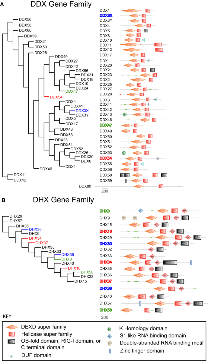
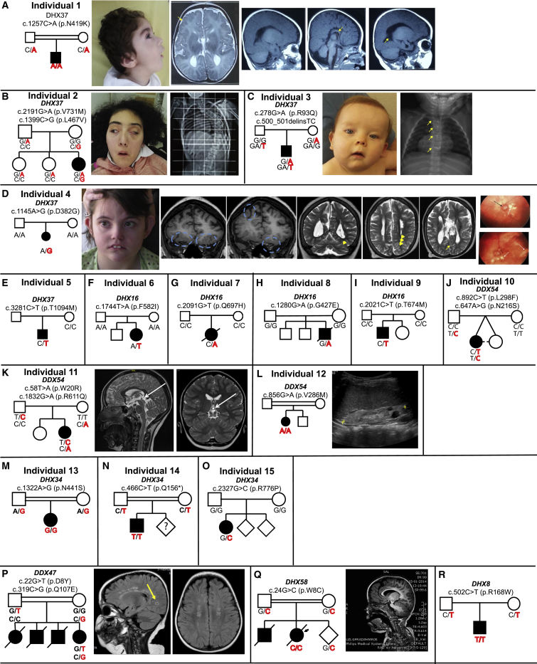
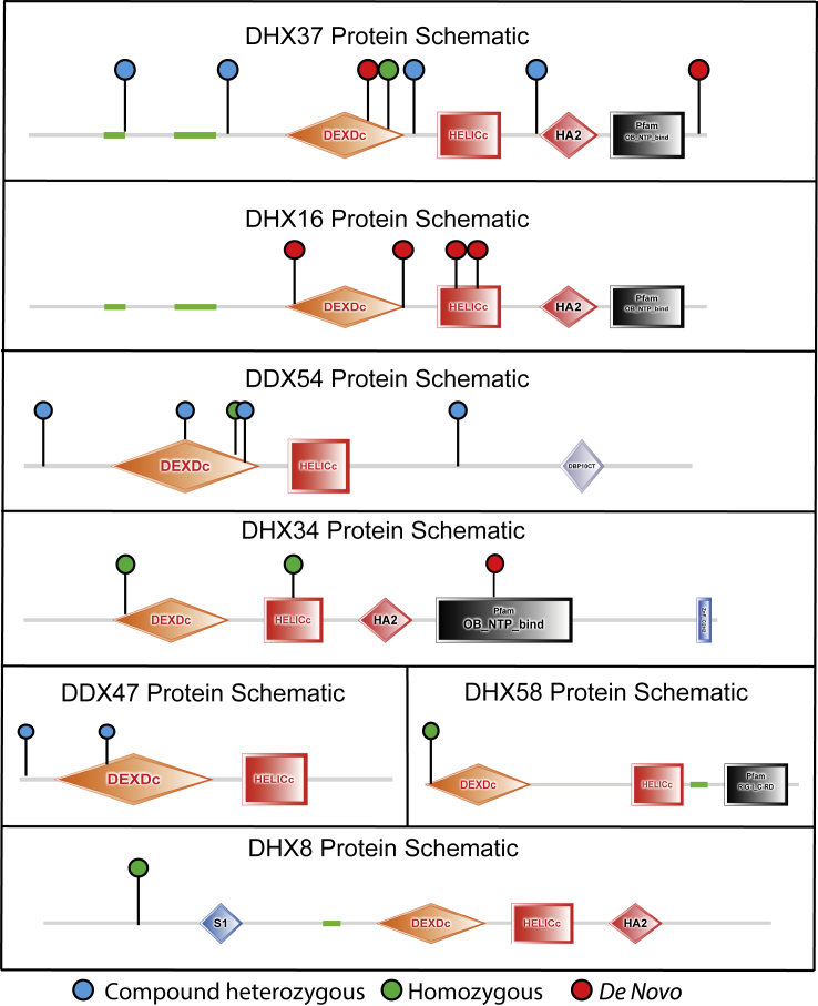
Members of a paralogous gene family in which variation in one gene is known to cause disease are eight times more likely to also be associated with human disease. Recent studies have elucidated DHX30 and DDX3X as genes for which pathogenic variant alleles are involved in neurodevelopmental disorders. We hypothesized that variants in paralogous genes encoding members of the DExD/H-box RNA helicase superfamily might also underlie developmental delay and/or intellectual disability (DD and/or ID) disease phenotypes. Here we describe 15 unrelated individuals who have DD and/or ID, central nervous system (CNS) dysfunction, vertebral anomalies, and dysmorphic features and were found to have probably damaging variants in DExD/H-box RNA helicase genes. In addition, these individuals exhibit a variety of other tissue and organ system involvement including ocular, outer ear, hearing, cardiac, and kidney tissues. Five individuals with homozygous (one), compound-heterozygous (two), or de novo (two) missense variants in DHX37 were identified by exome sequencing. We identified ten total individuals with missense variants in three other DDX/DHX paralogs: DHX16 (four individuals), DDX54 (three individuals), and DHX34 (three individuals). Most identified variants are rare, predicted to be damaging, and occur at conserved amino acid residues. Taken together, these 15 individuals implicate the DExD/H-box helicases in both dominantly and recessively inherited neurodevelopmental phenotypes and highlight the potential for more than one disease mechanism underlying these disorders.

Keywords: DExD/H-box RNA helicase family; developmental delay; human paralogs; intellectual disability.


Conflict of interest statement

J.R.L. serves on the scientific advisory board for Baylor Genetics. J.R.L. has stock ownership in 23andMe, is a paid consultant for Regeneron Pharmaceuticals, and is a coinventor on multiple US and European patents related to molecular diagnostics for inherited neuropathies, eye diseases, and bacterial genomic fingerprinting.

Figures

Figure 1
Figure 1
DExD/H-Box RNA Helicase Protein Family (A and B) Phylogeny trees for DEAD-box (A, left) and DEAH-box (B, left) genes were generated with Phylogeny.fr. The trees are presented with branch length ignored to facilitate clarity. Protein schematics for each DEAD-box (A, right) and DEAH-box (B, right) gene family are shown with known functional domains indicated. Genes that have been previously associated with human disease are indicated in blue text. Gene family members for which evidence is presented in this manuscript are indicated in red text. Gene family members for which only one individual is presented in this manuscript are indicated in green text.
Figure 2
Figure 2
Individual Pedigrees and Clinical Findings (A–O) Pedigrees, genotypes, and available consented clinical images are shown for individuals 1–15. (A) Individual 1: arrows indicate polymicrogyria, dysgenesis of the corpus callosum, and cerebellar volume loss. (B) Individual 2: an x-ray image shows scoliosis. (C) Individual 3: arrows indicate segmentation anomalies of the vertebra. (D) Individual 4: circles indicate polymicrogyria, arrowheads indicate polymicrogyria, the arrow indicates a cyst, white arrows indicate chorioretinal lacunae, and black arrows indicate optic nerve colobomas. (K) Individual 11: white arrows indicate pineal gland cysts. (L) Individual 12: ultrasound images show polycystic kidneys. (P–R) Additional candidate variants were identified in single families for three genes, DDX47 (MIM: 615428), DHX58 (MIM: 608588), and DHX8 (MIM: 600396), and these are indicated at the bottom of the figure. (P) The MRI images from the individual with a candidate DDX47 variant show occipital infarct (yellow arrow) and white matter abnormalities. (Q)The MRI image from the individual with a candidate DHX58 variant shows a thin corpus callosum.
Figure 3
Figure 3
Variant Location Schematic Protein schematics showing the functional domains of the seven candidate genes with the location of the variants indicated on each. A red lollipop indicates a de novo variant, a blue lollipop indicates compound heterozygosity for variants, and a green lollipop indicates homozygosity for the variant.
Figure 4
Figure 4
Variant Inheritance Confirmation Phasing of DDX54 variant c.892C>T (p.Leu298Phe) (Chr12: 113,612,724_G>A, paternally inherited) and c.647A>G (p.Asn216Ser) (Chr12: 113,614,866_T>C, de novo). (A) Dideoxy Sanger sequencing of two clones generated from long-range PCR amplification of both variants demonstrates one clone containing neither variant and one clone containing both variants, consistent with an in cis variant allele configuration. (B) Droplet digital PCR probes were designed with specificity to each variant, and optimization demonstrates that both variants are detected in individual 10, but not in a wild-type control sample. (C) Two-dimensional droplet distribution demonstrates dense clustering in the +/+ cluster, indicating co-segregation of both variants within droplets. (D and E) This clustering is not observed when genomic DNA is treated with SphI digestion prior to ddPCR. An SphI restriction site is located between the variants, and digestion leads to independent segregation of variants within droplets.

References

    1. Barshir R., Hekselman I., Shemesh N., Sharon M., Novack L., Yeger-Lotem E. Role of duplicate genes in determining the tissue-selectivity of hereditary diseases. PLoS Genet. 2018;14:e1007327. - PMC - PubMed
    1. Beighton P., De Paepe A., Steinmann B., Tsipouras P., Wenstrup R.J., Ehlers-Danlos National Foundation (USA) and Ehlers-Danlos Support Group (UK) Ehlers-Danlos syndromes: Revised nosology, Villefranche, 1997. Am. J. Med. Genet. 1998;77:31–37. - PubMed
    1. Malfait F., Francomano C., Byers P., Belmont J., Berglund B., Black J., Bloom L., Bowen J.M., Brady A.F., Burrows N.P. The 2017 international classification of the Ehlers-Danlos syndromes. Am. J. Med. Genet. C. Semin. Med. Genet. 2017;175:8–26. - PubMed
    1. Gonzaga-Jauregui C., Gamble C.N., Yuan B., Penney S., Jhangiani S., Muzny D.M., Gibbs R.A., Lupski J.R., Hecht J.T. Mutations in COL27A1 cause Steel syndrome and suggest a founder mutation effect in the Puerto Rican population. Eur. J. Hum. Genet. 2015;23:342–346. - PMC - PubMed
    1. Yamamoto S., Jaiswal M., Charng W.L., Gambin T., Karaca E., Mirzaa G., Wiszniewski W., Sandoval H., Haelterman N.A., Xiong B. A drosophila genetic resource of mutants to study mechanisms underlying human genetic diseases. Cell. 2014;159:200–214. - PMC - PubMed

Publication types