. 2019 Oct 15;70(19):5245-5258.
doi: 10.1093/jxb/erz305.

PPR-SMR1 is required for the splicing of multiple mitochondrial introns, interacts with Zm-mCSF1, and is essential for seed development in maize

Affiliations

PPR-SMR1 is required for the splicing of multiple mitochondrial introns, interacts with Zm-mCSF1, and is essential for seed development in maize

Zongliang Chen et al. J Exp Bot. .

Abstract

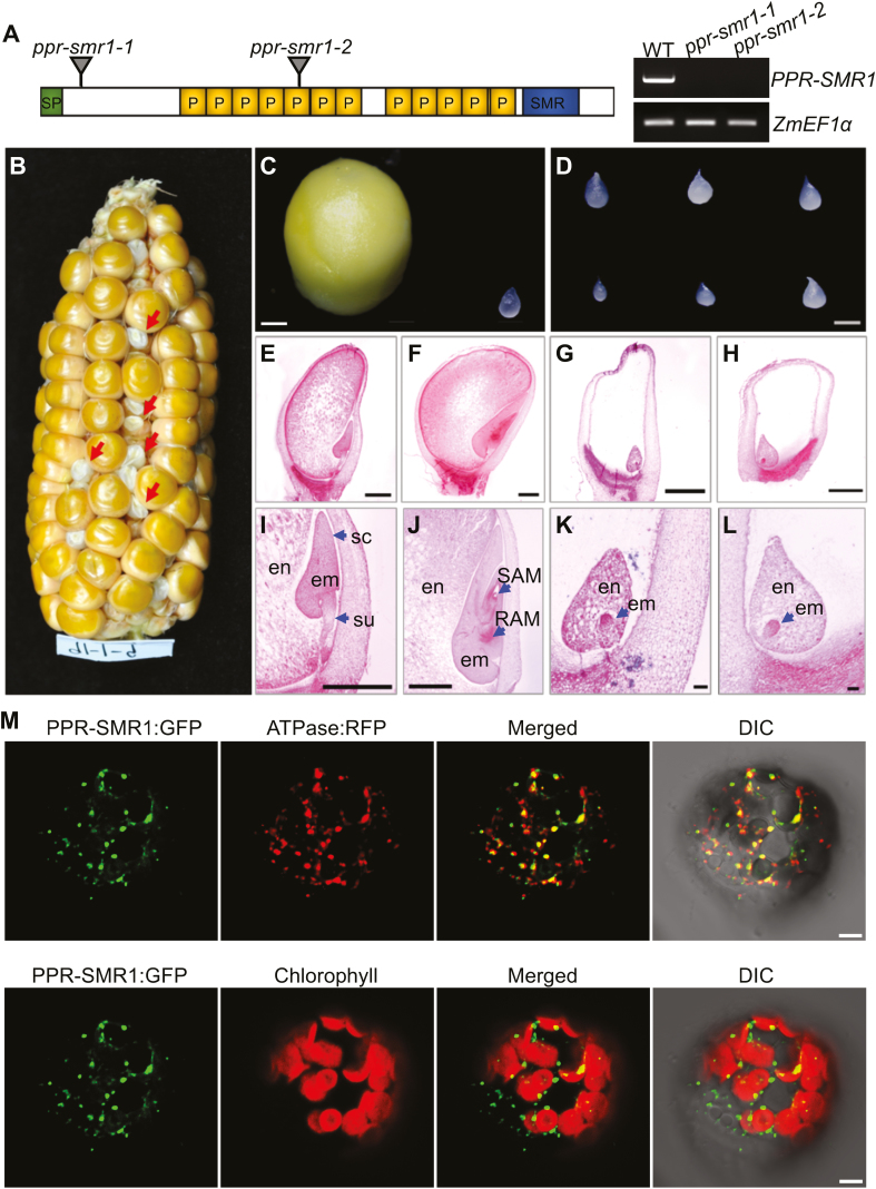
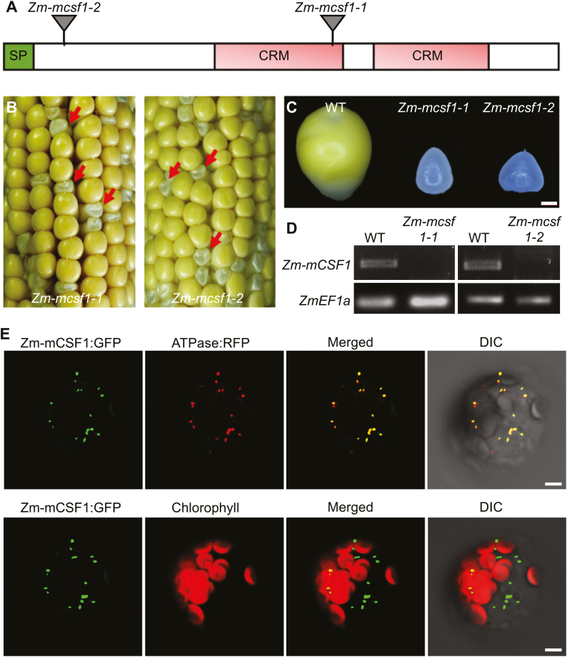
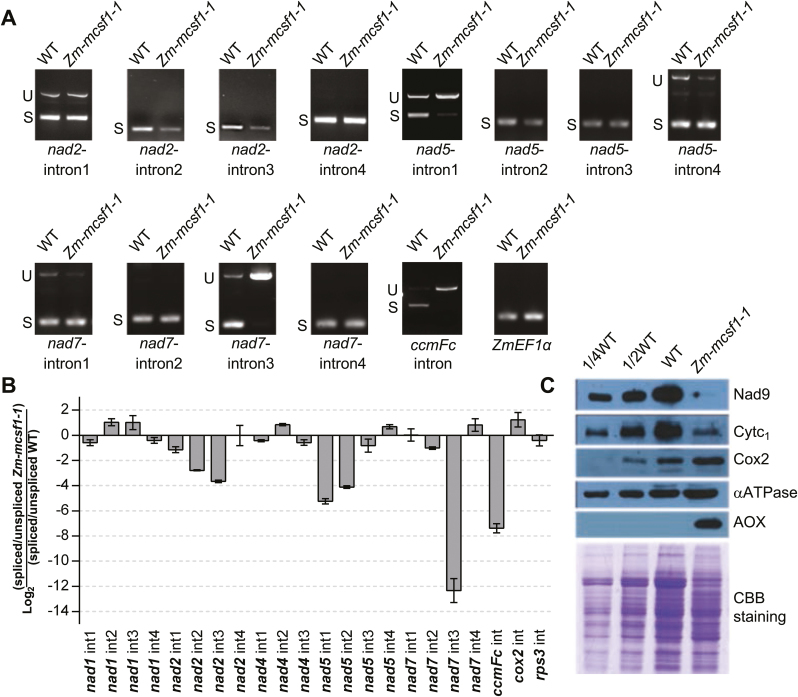
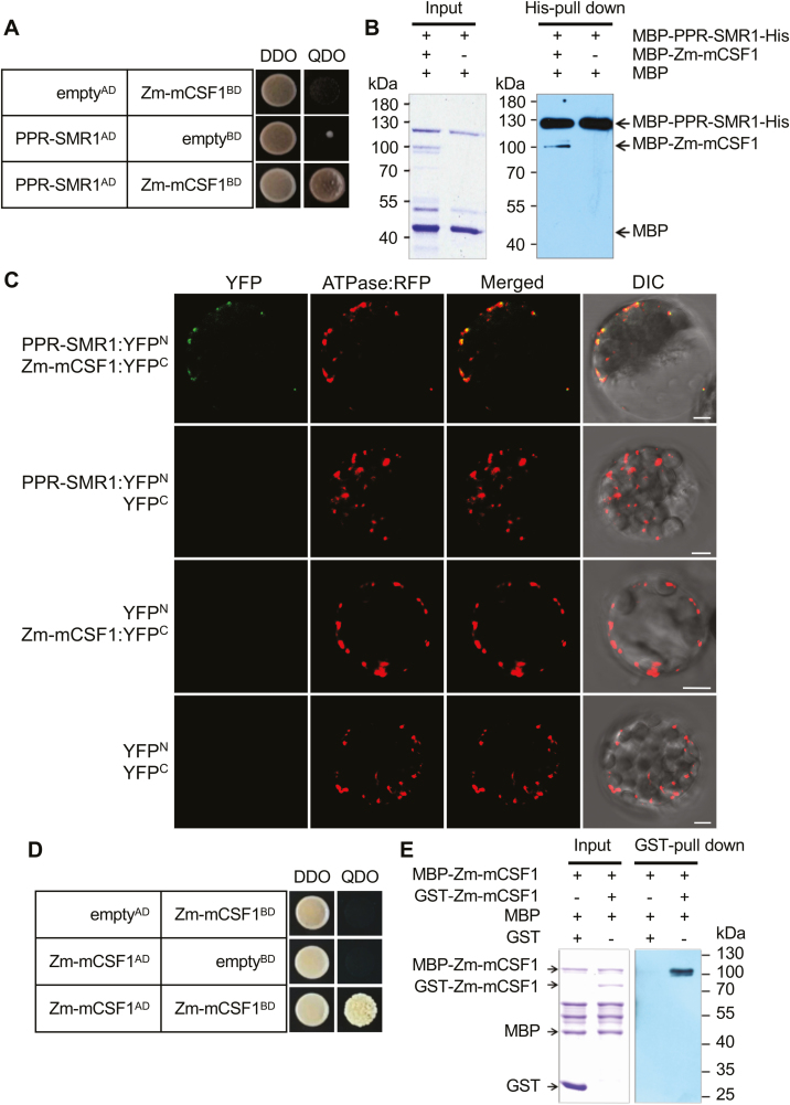
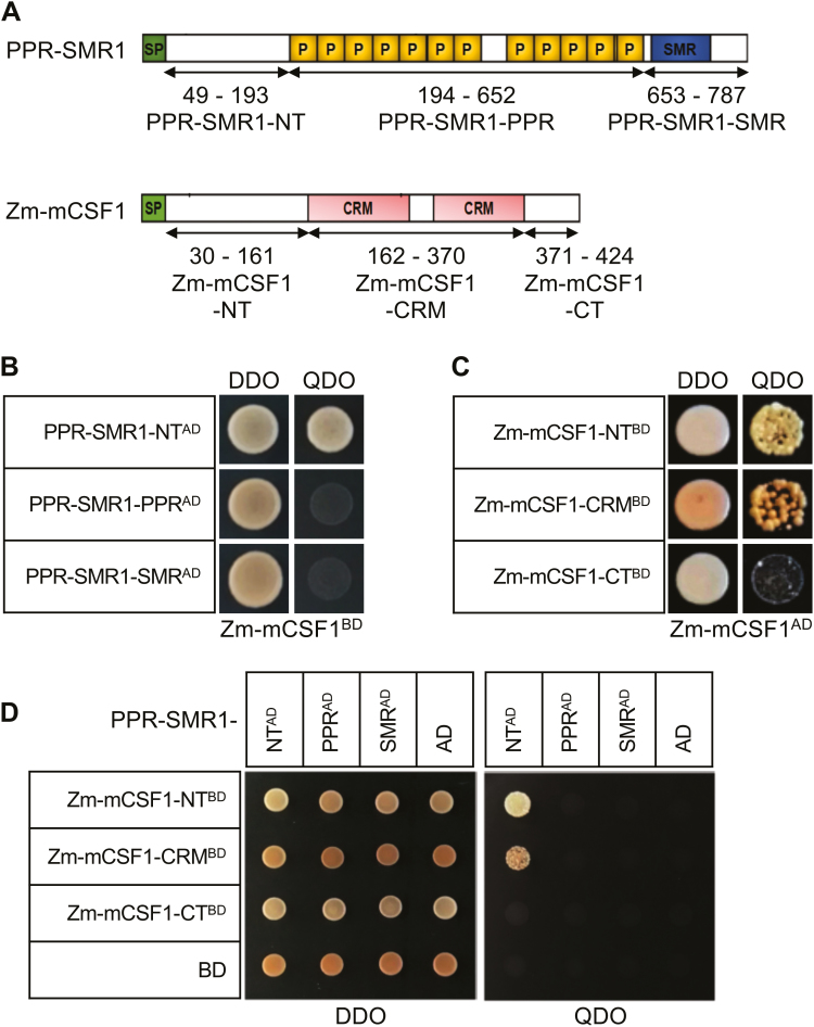
Group II introns are ribozymes that can excise themselves from precursor-RNA transcripts, but plant organellar group II introns have structural deviations that inhibit ribozyme activity. Therefore, splicing of these introns requires the assistance of nuclear- and/or organellar-encoded splicing factors; however, how these splicing factors function remains unclear. In this study, we report the functions and interactions of two splicing factors, PPR-SMR1 and Zm-mCSF1, in intron splicing in maize mitochondria. PPR-SMR1 is a SMR domain-containing pentatricopeptide repeat (PPR) protein and Zm-mCSF1 is a CRM domain-containing protein, and both are targeted to mitochondria. Loss-of-function mutations in each of them severely arrests embryogenesis and endosperm development in maize. Functional analyses indicate that PPR-SMR1 and Zm-mCSF1 are required for the splicing of most mitochondrial group II introns. Among them, nad2-intron 2 and 3, and nad5-intron 1 are PPR-SMR1/Zm-mCSF1-dependent introns. Protein interaction assays suggest that PPR-SMR1 can interact with Zm-mCSF1 through its N-terminus, and that Zm-mCSF1 is self-interacting. Our findings suggest that PPR-SMR1, a novel splicing factor, acts in the splicing of multiple group II introns in maize mitochondria, and the protein-protein interaction between it and Zm-mCSF1 might allow the formation of large macromolecular splicing complexes.

Keywords: Group II introns; maize; mitochondria; organelle biogenesis; pentatricopeptide repeat (PPR) proteins; seed development.


Figures

Fig. 1.
Fig. 1.
Overview of maize PPR-SMR1. (A) Diagram of the PPR-SMR1 protein containing 12 PPR domains, with the positions of the Mutator insertions indicated. Expression of PPR-SMR1 is absent from ppr-smr1 mutants. SP, signaling peptide; P, PPR motif; SMR, small MutS-related. (B) A maize ear showing 3:1 segregation for wild-type (WT) and ppr-smr1-1 mutant kernels (arrows). (C) Dissection of WT (left) and ppr-smr1-1 (right) endosperm at 11 d after pollination (DAP). (D) Mutant endosperm dissected from reciprocal crosses of ppr-smr1-1 and ppr-smr1-2 kernels. (E–L) Comparisons of WT and ppr-smr1-1 kernel development at 9 DAP and 13 DAP. WT kernels at 9 DAP (E, I) and 13 DAP (F, J); ppr-smr1-1 kernels at 9 DAP (G, K) and 13 DAP (H, L). en, endosperm; em, embryo; su, suspensor; sc, scutellum; RAM, root apical meristem; SAM, shoot apical meristem. (M) Localization of the PPR-SMR1 protein. Mesophyll protoplasts from Arabidopsis were transformed with PPR-SMR1::GFP and ATPase::RFP constructs and imaged using confocal microscopy. In the upper images, mitochondria are labeled by fluorescence of ATPase::RFP, and in the lower images, chloroplasts are marked by autofluorescence. GFP, green fluorescent protein; DIC, differential interference contrast. Scale bars are 1 mm in (C–J), 0.1 mm in (K, L), and 5 µm in (M).
Fig. 2.
Fig. 2.
PPR-SMR1 is involved in splicing of maize group II introns. (A) Inefficient splicing of group II introns of nad1, nad2, nad4, nad5, and nad7 in the embryos and endosperms of ppr-smr1-1 mutants as determined by reverse-transcription PCR (RT-PCR). S, introns are spliced; U, introns are retained. (B) Quantitative real-time RT-PCR (qRT-PCR) analysis of the 22 intron-containing mitochondrial transcripts in ppr-smr1 mutants. Total RNA was extracted from embryos an endosperms of ppr-smr1 mutants and their wild-type siblings at 12 d after pollination. ZmEF1α was used to normalize the quantifications. Data are means (±SD) of three biological replicates. (C) Immunoblot analysis of core subunits of mitochondrial complexes in ppr-smr1-1 mutants. Immunoblots of embryo and endosperm extract (20 µg protein or the indicated dilutions) were probed with antibodies specific for subunits of Complex I (Nad9), Complex III (Cytochrome c1, Cytc1), Complex IV (Cox2), and ATP synthase (αATPase). AOX, alternative oxidase. Total protein input can be seen on the Ponceau S-stained blots below. (This figure is available in color at JXB online.)
Fig. 3.
Fig. 3.
Overview of Zm-mCSF1. (A) Diagram of the Zm-mCSF1 protein containing two CRS1-YhbY domains (CRM) with the positions of the Mutator insertions in alleles indicated. SP, signaling peptide. (B) Seed phenotype of Zm-mcsf1 mutants. Ears of Zm-mcsf1-1 and Zm-mcsf1-2 were harvested at 15 d after pollination. The arrows indicate homozygous Zm-mcsf1 seed mutants classified as empty pericarp (emp). (C) Comparison of endosperm morphologies of Zm-mcsf1-1 and Zm-mcsf1-2 with the wild-type (WT). (D) Reverse-transcription PCR demonstrating the absence of Zm-mCSF1 mRNA in the Zm-mcsf1 mutants. (E) Localization of the Zm-mCSF1 protein. Mesophyll protoplasts from Arabidopsis were transformed with Zm-mCSF1::GFP and ATPase::RFP constructs and imaged using confocal microscopy. In the upper images, mitochondria are labeled by fluorescence of ATPase::RFP, and in the lower images, chloroplasts are marked by autofluorescence. DIC, differential interference contrast. Scale bars are 1 mm in (C) and 5 µm in (E).
Fig. 4.
Fig. 4.
Zm-mCSF1 is involved in splicing of maize group II introns. (A) Inefficient splicing of group II introns of nad2, nad5, nad7, and ccmFC in the endosperm of Zm-mcsf1-1 mutants by reverse-transcription PCR (RT-PCR). S, introns are spliced; U, introns are retained. (B) qRT-PCR analysis of the 22 intron-containing mitochondrial transcripts in Zm-mcsf1-1 mutants. RNA was extracted from the embryos and endosperms of Zm-mcsf1-1 mutants and their wild-type siblings at 12 d after pollination. ZmEF1α was used to normalize the quantifications. Data are means (±SD) of three biological replicates. (C) Immunoblot analysis of core subunits of mitochondrial complexes in Zm-mcsf1-1 mutants. Immunoblots of embryos and endosperms extract (20 µg protein or the indicated dilutions) were probed with antibodies specific for subunits of Complex I (Nad9), Complex III (Cytochrome c1, Cytc1), Complex IV (Cox2), and ATP synthase (αATPase). AOX, alternative oxidase. Total protein input can be seen in the Coomassie Brilliant Blue (CBB) staining below. (This figure is available in color at JXB online.)
Fig. 5.
Fig. 5.
PPR-SMR1 protein interacts with Zm-mCSF1. (A) Yeast two-hybrid (Y2H) analysis of PPR-SMR1 and Zm-mCSF1 interaction. The Y2HGold strain harboring the indicated bait and prey constructs were spotted on synthetic dropout (SD) medium without Leu and Trp (double-dropout, DDO) and SD without Ade, Leu, Trp, and His (quadruple-dropout, QDO). Yeast cultures on DDO control plates demonstrate the existence of both plasmids. Positive interactions were verified by growth on QDO plates. (B) Recombinant protein MBP-PPR-SMR1-His interacts with MBP-Zm-mCSF1 as determined using in vitro His pull-down assays. (C) Bimolecular fluorescence complementation (BiFC) analysis of the interaction of PPR-SMR1 and Zm-mCSF1 in mesophyll protoplasts of Arabidopsis. Yellow fluorescent protein (YFP) was split into N- and C-terminus, and PPR-SMR1 was fused with the N-terminus of YFP and Zm-mCSF1 was fused with C-terminus of YFP. Scale bars are 5 µm. (D) Zm-mCSF1AD physically interacts with Zm-mCSF1BD in Y2H assays. Yeast Y2HGold was transformed with paired constructs Zm-mCSF1AD and Zm-mCSF1BD, and AD (activating domain) and BD (binding domain) were used as negative controls. (E) Recombinant protein MBP-Zm-mCSF1 interacts with GST-Zm-mCSF1 as determined using in vitro glutathione S-transferase (GST) pull-down assays. ‘+’ and ‘−’ indicate the presence and absence of the corresponding proteins in the reactions, respectively.
Fig. 6.
Fig. 6.
Protein fragments required for interactions of maize PPR-SMR1 and Zm-mCSF1. (A) Schematic diagram of full-length and split fragment fusions of PPR-SMR1 and Zm-mCSF1 to the GAL4 activating domain (AD) or the GAL4 DNA binding domain (BD), respectively. The PPR-SMR1 protein is split into three fragments: PPR-SMR1-NT (N-terminus, 49–193), PPR-SMR1-PPR (194–652), and PPR-SMR1-SMR (653–787). The Zm-mCSF1 protein is split into three fragments: Zm-mCSF1-NT (N-terminus, 30–161), Zm-mCSF1-CRM (162–370), and Zm-mCSF1-CT (371–424). (B–D) Yeast two-hybrid (Y2H) assays demonstrating the physical interactions. The Y2HGold harboring the indicated bait and prey constructs were spotted on synthetic dropout (SD) medium without Leu and Trp (double-dropout, DDO) and SD without Ade, Leu, Trp, and His (quadruple-dropout, QDO). (B) PPR-SMR1-NTAD physically interacts with Zm-mCSF1BD. (C) Zm-mCSF1-NTBD and Zm-mCSF1-CRMBD interact with Zm-mCSF1AD. (D) PPR-SMR1-NTAD physically interacts with Zm-mCSF1-NTBD and Zm-mCSF1-CRMBD. (This figure is available in color at JXB online.)

References

    1. Asakura Y, Barkan A. 2007. A CRM domain protein functions dually in group I and group II intron splicing in land plant chloroplasts. The Plant Cell 19, 3864–3875. - PMC - PubMed
    1. Asakura Y, Bayraktar OA, Barkan A. 2008. Two CRM protein subfamilies cooperate in the splicing of group IIB introns in chloroplasts. RNA 14, 2319–2332. - PMC - PubMed
    1. Barkan A, Klipcan L, Ostersetzer O, Kawamura T, Asakura Y, Watkins KP. 2007. The CRM domain: an RNA binding module derived from an ancient ribosome-associated protein. RNA 13, 55–64. - PMC - PubMed
    1. Barkan A, Small I. 2014. Pentatricopeptide repeat proteins in plants. Annual Review of Plant Biology 65, 415–442. - PubMed
    1. Bonen L. 2008. Cis- and trans-splicing of group II introns in plant mitochondria. Mitochondrion 8, 26–34. - PubMed

Publication types