

Astragaloside IV inhibits oxidized low‑density lipoprotein‑induced endothelial damage via upregulation of miR‑140‑3p
- PMID: 31257467
- PMCID: PMC6657972
- DOI: 10.3892/ijmm.2019.4257
Astragaloside IV inhibits oxidized low‑density lipoprotein‑induced endothelial damage via upregulation of miR‑140‑3p
Retraction in
-
[Retracted] Astragaloside IV inhibits oxidized low‑density lipoprotein‑induced endothelial damage via upregulation of miR‑140‑3p.Int J Mol Med. 2023 Feb;51(2):17. doi: 10.3892/ijmm.2023.5220. Epub 2023 Jan 20. Int J Mol Med. 2023. PMID: 36660951 Free PMC article.
Abstract
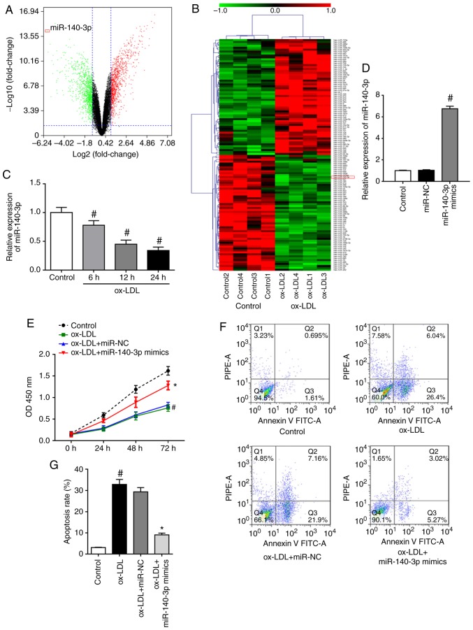
Oxidized low‑density lipoprotein (ox‑LDL)‑mediated endothelial cell injury has an important role in the vascular complications of type 2 diabetes. Astragaloside IV (ASV) is an active component of Radix Astragali, which has been demonstrated to exert protective effects against endothelial damage. The present study explored whether microRNAs (miRNAs) are involved in mediating the protective effects of ASV on ox‑LDL‑induced damage in human umbilical vein endothelial cells (HUVECs). RNA sequencing and reverse transcription‑quantitative PCR analyses revealed that ox‑LDL treatment significantly downregulated miR‑140‑3p expression in HUVECs. miR‑140‑3p overexpression promoted cell proliferation and inhibited apoptosis in ox‑LDL‑induced HUVECs. However, inhibition of miR‑140‑3p expression could reverse the effects of ASV on ox‑LDL‑induced HUVECs and reactivate ASV‑inhibited PI3K/Akt signaling in ox‑LDL‑induced HUVECs. In addition, Krüppel‑like factor 4 (KLF4) was identified as a target of miR‑140‑3p in ox‑LDL‑treated HUVECs. Subsequent experiments revealed that KLF4 overexpression partially counteracted the protective effects of miR‑140‑3p or ASV treatment in ox‑LDL‑induced HUVECs. Taken together, the current findings demonstrated that the protective effects of ASV on HUVECs were dependent on miR‑140‑3p upregulation and subsequent inhibition of KLF4 expression, which in turn suppressed the PI3K/Akt signaling pathway. The present results shed light to the molecular mechanism by which ASV alleviated ox‑LDL‑induced endothelial cell damage.
Figures


Similar articles
-
Salidroside attenuates oxidized low‑density lipoprotein‑induced endothelial cell injury via promotion of the AMPK/SIRT1 pathway.Int J Mol Med. 2019 Jun;43(6):2279-2290. doi: 10.3892/ijmm.2019.4153. Epub 2019 Apr 1. Int J Mol Med. 2019. PMID: 30942428 Free PMC article.
-
Oxymatrine attenuates oxidized low‑density lipoprotein‑induced HUVEC injury by inhibiting NLRP3 inflammasome‑mediated pyroptosis via the activation of the SIRT1/Nrf2 signaling pathway.Int J Mol Med. 2021 Oct;48(4):187. doi: 10.3892/ijmm.2021.5020. Epub 2021 Aug 9. Int J Mol Med. 2021. PMID: 34368883 Free PMC article.
-
Downregulation of microRNA‑34a inhibits oxidized low‑density lipoprotein‑induced apoptosis and oxidative stress in human umbilical vein endothelial cells.Int J Mol Med. 2018 Aug;42(2):1134-1144. doi: 10.3892/ijmm.2018.3663. Epub 2018 May 9. Int J Mol Med. 2018. PMID: 29750293
-
Rosuvastatin protects against oxidized low‑density lipoprotein‑induced endothelial cell injury of atherosclerosis in vitro.Mol Med Rep. 2019 Jan;19(1):432-440. doi: 10.3892/mmr.2018.9666. Epub 2018 Nov 19. Mol Med Rep. 2019. PMID: 30483737 Free PMC article.
-
miR‑21/PTEN pathway mediates the cardioprotection of geniposide against oxidized low‑density lipoprotein‑induced endothelial injury via suppressing oxidative stress and inflammatory response.Int J Mol Med. 2020 May;45(5):1305-1316. doi: 10.3892/ijmm.2020.4520. Epub 2020 Feb 28. Int J Mol Med. 2020. PMID: 32323738 Free PMC article.
Cited by
-
The Molecular Basis of the Anti-Inflammatory Property of Astragaloside IV for the Treatment of Diabetes and Its Complications.Drug Des Devel Ther. 2023 Mar 10;17:771-790. doi: 10.2147/DDDT.S399423. eCollection 2023. Drug Des Devel Ther. 2023. PMID: 36925998 Free PMC article. Review.
-
Circ_0068087 Silencing Ameliorates Oxidized Low-Density Lipoprotein-Induced Dysfunction in Vascular Endothelial Cells Depending on miR-186-5p-Mediated Regulation of Roundabout Guidance Receptor 1.Front Cardiovasc Med. 2021 May 26;8:650374. doi: 10.3389/fcvm.2021.650374. eCollection 2021. Front Cardiovasc Med. 2021. PMID: 34124191 Free PMC article.
-
Astragaloside IV protects ATDC5 cells from lipopolysaccharide-caused damage through regulating miR-203/MyD88.Pharm Biol. 2020 Dec;58(1):89-97. doi: 10.1080/13880209.2019.1705355. Pharm Biol. 2020. PMID: 31906765 Free PMC article.
-
Isoflavones from Semen Sojae Preparatum Improve Atherosclerosis and Oxidative Stress by Modulating Nrf2 Signaling Pathway through Estrogen-Like Effects.Evid Based Complement Alternat Med. 2022 Apr 7;2022:4242099. doi: 10.1155/2022/4242099. eCollection 2022. Evid Based Complement Alternat Med. 2022. PMID: 35432565 Free PMC article.
-
Transcriptomic Analysis Reveals the Protection of Astragaloside IV against Diabetic Nephropathy by Modulating Inflammation.Oxid Med Cell Longev. 2020 Aug 12;2020:9542165. doi: 10.1155/2020/9542165. eCollection 2020. Oxid Med Cell Longev. 2020. PMID: 32855769 Free PMC article.
References
-
- Shore AC, Colhoun HM, Natali A, Palombo C, Khan F, Östling G, Aizawa K, Kennbäck C, Casanova F, Persson M, et al. Use of vascular assessments and novel biomarkers to predict cardiovascular events in type 2 Diabetes: The SUMMIT VIP study. Diabetes Care. 2018;41:2212–2219. doi: 10.2337/dc18-0185. - DOI - PubMed
