. 2019 Aug;20(2):1605-1612.
doi: 10.3892/mmr.2019.10401. Epub 2019 Jun 20.

Carvedilol improves liver cirrhosis in rats by inhibiting hepatic stellate cell activation, proliferation, invasion and collagen synthesis

Affiliations

Carvedilol improves liver cirrhosis in rats by inhibiting hepatic stellate cell activation, proliferation, invasion and collagen synthesis

Liping Ling et al. Mol Med Rep. 2019 Aug.

Abstract

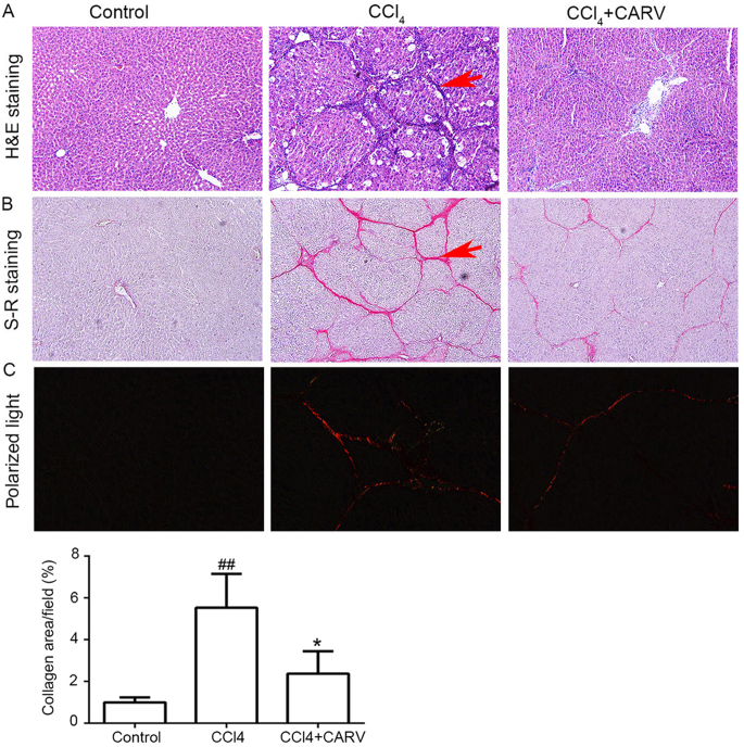
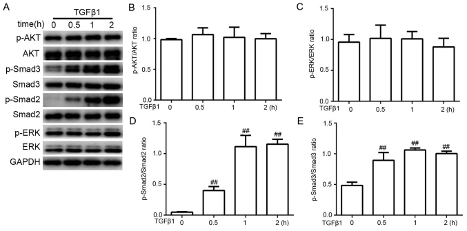
Portal hypertension (PHT) is one of the most severe consequences of liver cirrhosis. Carvedilol is a first‑line pharmacological treatment of PHT. However, the antifibrogenic effects of carvedilol on liver cirrhosis and the intrinsic mechanisms underlying these effects have not been thoroughly investigated. The present study aimed to investigate the antifibrogenic effects of carvedilol on liver cirrhosis in vivo and in vitro. Liver cirrhosis was induced in rats by carbon tetrachloride (CCl4) administration for 9 weeks; carvedilol was administered simultaneously in the experimental group. Blood samples were collected for serum biochemistry. Liver tissues were used for fibrosis evaluation, histological examination, immunohistochemistry and western blot analysis. The human hepatic stellate cell (HSC) line LX‑2 was used for in vitro studies. The effects of carvedilol on LX‑2 cell proliferation and invasion were evaluated by Cell Counting Kit‑8 assay and Transwell invasion assays, respectively. The effect of carvedilol on transforming growth factor β1 (TGFβ1)‑induced collagen synthesis in LX‑2 cells and the molecular mechanisms were examined by western blot analysis. The results demonstrated that carvedilol improved CCl4‑induced structural distortion and fibrosis in the liver. Carvedilol inhibited HSC activation, proliferation and invasion. Carvedilol inhibited HSC collagen synthesis through the TGFβ1/SMAD pathway. In conclusion, carvedilol may alleviate liver cirrhosis in rats by inhibiting HSC activation, proliferation, invasion and collagen synthesis. Carvedilol may be a potential treatment of early‑stage liver cirrhosis.


Figures

Figure 1.
Figure 1.
Carvedilol improves CCl4-induced structural distortion and fibrosis. (A) Representative histological images of H&E stained liver sections of non-cirrhotic Control rats, CCl4-intoxicatd rats and CCl4-intoxicatd rats treated with carvedilol; magnification, ×100. Arrow indicates the regenerative nodule. (B) Images of S-R staining of livers from rats in each group; magnification, ×100. Arrow indicates the collagen deposition. (C) Quantification of collagen-positive area in liver sections by polarized light microscopy; magnification, ×200. ##P<0.01 vs. control group; *P<0.05 vs. CCl4-intoxicated group. CARV, carvedilol; CCl4, carbon tetrachloride; H&E, hematoxylin and eosin; S-R, sirius red.
Figure 2.
Figure 2.
Carvedilol inhibits hepatic stellate cell activation in vivo and in vitro. (A) Representative images of immunohistochemical staining of α-SMA of liver sections of non-cirrhotic Control rats, CCl4-intoxicatd rats and CCl4-intoxicatd rats treated with carvedilol; magnification, ×100. (B) Protein expression levels of α-SMA in liver tissues were quantified by western blotting and densitometric analysis. (C) α-SMA protein expression levels in LX-2 cells treated with various concentrations of carvedilol. ##P<0.01 vs. control; **P<0.01 vs. CCl4-intoxicated group. α-SMA, α-smooth muscle actin; CARV, carvedilol; CCl4, carbon tetrachloride.
Figure 3.
Figure 3.
Carvedilol inhibits proliferative and invasive abilities of hepatic stellate cells. (A) Effects of a range of carvedilol concentrations on the proliferation of LX-2 cells were evaluated by Cell Counting Kit-8 assay. (B) Effects of carvedilol on the invasive ability of LX-2 cells were examined by Transwell migration assay; magnification, ×100. ##P<0.01 vs. serum-free Control; *P<0.05 vs. 0 µM carvedilol + 10% FBS; **P<0.01 vs. 0 µM carvedilol + 10% FBS. CARV, carvedilol.
Figure 4.
Figure 4.
Carvedilol inhibits TGFβ1-induced collagen synthesis of hepatic stellate cells. (A) TGFβ1-induced Col I and FN protein expression in LX-2 cells. (B) Carvedilol inhibited TGFβ1-induced Col I and FN protein expression in LX-2 cells. #P<0.05 vs. control; ##P<0.01 vs. control; *P<0.05 vs. 0 µM carvedilol + TGFβ1; **P<0.01 vs. 0 µM carvedilol + TGFβ1. CARV, carvedilol; Col I, type I collagen; FN, fibronectin; TGFβ1, transforming growth factor β1.
Figure 5.
Figure 5.
AKT, ERK, SMAD2 and SMAD3 phosphorylation levels in TGFβ1-stimulated LX-2 cells. (A) Representative western blotting analysis of protein expressions and densitometric analysis of the phosphorylation levels of (B) AKT, (C) ERK, (D) SMAD2 and (E) SMAD3 following TGFβ1 stimulation. ##P<0.01 vs. control. AKT, protein kinase B; p, phosphorylated; TGFβ1, transforming growth factor β1.
Figure 6.
Figure 6.
Carvedilol inhibits TGFβ1-induced hepatic stellate cell collagen synthesis through the TGFβ/SMAD pathway. (A) Carvedilol suppressed the TGFβ1-induced upregulation of p-SMAD2. (B) Carvedilol attenuated the TGFβ1-induced upregulation of p-SMAD3. (C) SIS3 treatment decreased the TGFβ1-induced expression of Col I and FN. #P<0.05 vs. control; ##P<0.01 vs. control; *P<0.05 vs. 0 µM carvedilol + TGFβ1, **P<0.01 vs. 0 µM carvedilol + TGFβ1. CARV, carvedilol; Col I, type I collagen; FN, fibronectin; p, phosphorylated; TGFβ1, transforming growth factor β1.

References

    1. Fernandez M. Molecular pathophysiology of portal hypertension. Hepatology. 2015;61:1406–1415. doi: 10.1002/hep.27343. - DOI - PubMed
    1. Bosch J, Groszmann RJ, Shah VH. Evolution in the understanding of the pathophysiological basis of portal hypertension: How changes in paradigm are leading to successful new treatments. J Hepatol. 2015;62:S121–S130. doi: 10.1016/j.jhep.2015.01.003. - DOI - PMC - PubMed
    1. Reiberger T, Ferlitsch A, Payer BA, Pinter M, Homoncik M, Peck-Radosavljevic M, Vienna Hepatic Hemodynamic Lab Non-selective β-blockers improve the correlation of liver stiffness and portal pressure in advanced cirrhosis. J Gastroenterol. 2012;47:561–568. doi: 10.1007/s00535-011-0517-4. - DOI - PubMed
    1. Bosch J, Abraldes JG, Fernández M, García-Pagán JC. Hepatic endothelial dysfunction and abnormal angiogenesis: New targets in the treatment of portal hypertension. J Hepatol. 2010;53:558–567. doi: 10.1016/j.jhep.2010.03.021. - DOI - PubMed
    1. Iwakiri Y, Groszmann RJ. Vascular endothelial dysfunction in cirrhosis. J Hepatol. 2007;46:927–934. doi: 10.1016/j.jhep.2007.02.006. - DOI - PubMed

MeSH terms