

Dexmedetomidine protects aged rats from postoperative cognitive dysfunction by alleviating hippocampal inflammation
- PMID: 31257507
- PMCID: PMC6691222
- DOI: 10.3892/mmr.2019.10438
Dexmedetomidine protects aged rats from postoperative cognitive dysfunction by alleviating hippocampal inflammation
Abstract
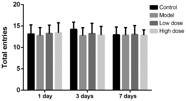
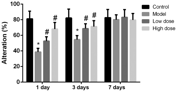
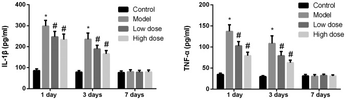
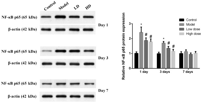
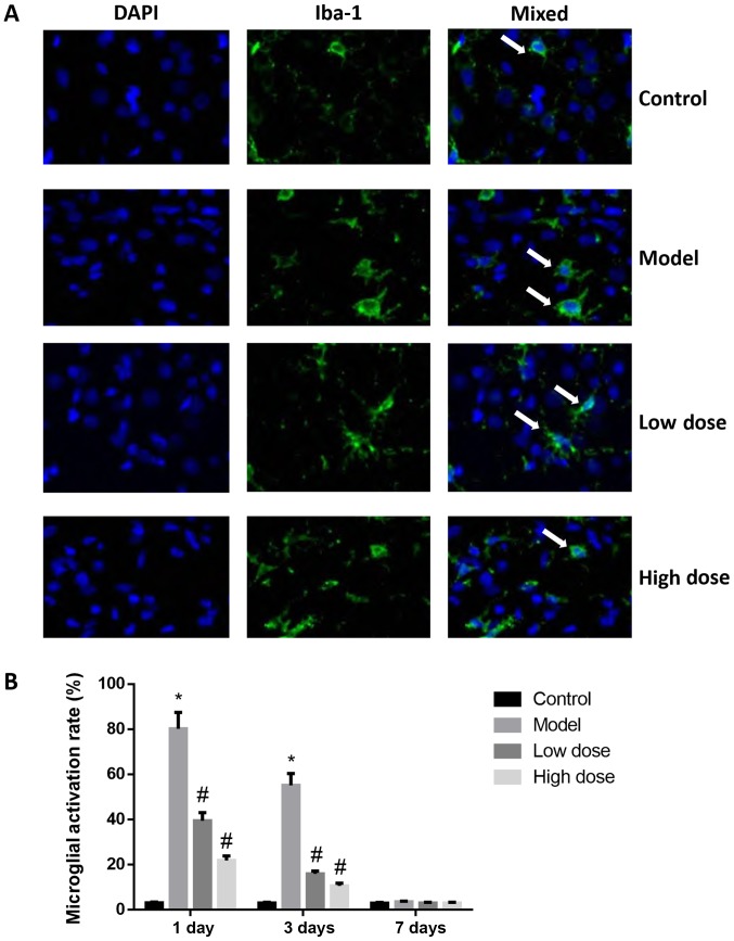
The present study investigated the effect of dexmedetomidine on hippocampal inflammation and cognitive function in rats with postoperative cognitive dysfunction (POCD). A total of 80 healthy male Sprague Dawley rats were used, 72 of which developed POCD. The rats were randomly divided into four groups: The control, model, low‑dose and high‑dose dexmedetomidine anesthesia groups. A POCD model was established and dexmedetomidine was administered. Cognitive function tests were performed and expression levels of interleukin 1β (IL‑1β), tumor necrosis factor‑α (TNF‑α) and NF‑κB biomarkers were evaluated on the first, third and seventh day following modeling. The cognitive function of rats was measured using a Y‑maze test. The expression levels of IL‑1β and TNF‑α in the hippocampus were determined by ELISA. The protein expression levels of NF‑κB p65 in the hippocampus were determined by western blotting. It was revealed that at 1, 3 and 7 days after surgery, there were no alterations in the exercise ability of rats in the different groups, as reflected by the number of rats passing the alternative arms in the Y‑maze. On the first and third day after surgery, the cognitive dysfunction reflected by the alteration scores of the low‑dose and high‑dose dexmedetomidine anesthesia groups were significantly higher than those of the model group, and the increase in the high‑dose group was more pronounced. Additionally, on the first day after surgery, the expression levels of IL‑1β, TNF‑α and NF‑κB in the hippocampi of rats in the low‑ and high‑dose dexmedetomidine anesthesia groups were significantly lower than those in the model group, and the decrease was more pronounced in the high‑dose group. At 7 days after surgery, the differences in expression levels of IL‑1β, TNF‑α and NF‑κB in the hippocampus among groups were not identified to be statistically significantly different. Taken together, the results of the present study indicated that dexmedetomidine may inhibit hippocampal inflammation induced by surgical trauma, and that dexmedetomidine may effectively improve postoperative cognitive function in rats.
Figures
Similar articles
-
Dexmedetomidine improves early postoperative cognitive dysfunction in aged mice.Eur J Pharmacol. 2015 Jan 5;746:206-12. doi: 10.1016/j.ejphar.2014.11.017. Epub 2014 Nov 20. Eur J Pharmacol. 2015. PMID: 25460022
-
[Dexmedetomidine alleviates postoperative cognitive dysfunction in aged rats probably via silent information regulator 1 pathway].Nan Fang Yi Ke Da Xue Xue Bao. 2018 Aug 30;38(9):1071-1075. doi: 10.12122/j.issn.1673-4254.2018.09.08. Nan Fang Yi Ke Da Xue Xue Bao. 2018. PMID: 30377100 Free PMC article. Chinese.
-
Effect of dexmedetomidine on rats with convulsive status epilepticus and association with activation of cholinergic anti-inflammatory pathway.Biochem Biophys Res Commun. 2018 Jan 1;495(1):421-426. doi: 10.1016/j.bbrc.2017.10.124. Epub 2017 Nov 11. Biochem Biophys Res Commun. 2018. PMID: 29080744
-
Effect of dexmedetomidine on postoperative cognitive dysfunction and inflammation in patients after general anaesthesia: A PRISMA-compliant systematic review and meta-analysis.Medicine (Baltimore). 2019 May;98(18):e15383. doi: 10.1097/MD.0000000000015383. Medicine (Baltimore). 2019. PMID: 31045788 Free PMC article.
-
The use of dexmedetomidine in anesthesia and intensive care: a review.Curr Pharm Des. 2012;18(38):6257-65. doi: 10.2174/138161212803832272. Curr Pharm Des. 2012. PMID: 22762468 Review.
Cited by
-
Nanopathways modulating postoperative cognitive dysfunction: extracellular vesicles.Front Cell Dev Biol. 2025 Jun 30;13:1613378. doi: 10.3389/fcell.2025.1613378. eCollection 2025. Front Cell Dev Biol. 2025. PMID: 40661144 Free PMC article. Review.
-
The Influence of Different Dexmedetomidine Doses on Cognitive Function at Early Period of Patients Undergoing Laparoscopic Extensive Total Hysterectomy.J Healthc Eng. 2021 Sep 28;2021:3531199. doi: 10.1155/2021/3531199. eCollection 2021. J Healthc Eng. 2021. Retraction in: J Healthc Eng. 2022 Nov 25;2022:9878490. doi: 10.1155/2022/9878490. PMID: 34621501 Free PMC article. Retracted.
-
Goal-directed therapy based on rScO2 monitoring in elderly patients with one-lung ventilation: a randomized trial on perioperative inflammation and postoperative delirium.Trials. 2022 Aug 19;23(1):687. doi: 10.1186/s13063-022-06654-6. Trials. 2022. PMID: 35986421 Free PMC article. Clinical Trial.
-
Association between estimated glomerular filtration rate and memory function mediated by brain Gray matter volumes: a cross-sectional study using the ADNI database.BMC Geriatr. 2025 Aug 15;25(1):628. doi: 10.1186/s12877-025-06326-5. BMC Geriatr. 2025. PMID: 40817206 Free PMC article.
-
The effects of dexmedetomidine on postoperative cognitive dysfunction in rats with bone fractures undergoing open reduction.Am J Transl Res. 2024 Jul 15;16(7):3005-3013. doi: 10.62347/QQKB3082. eCollection 2024. Am J Transl Res. 2024. PMID: 39114713 Free PMC article.
References
-
- Berger M, Nadler JW, Browndyke J, Terrando N, Ponnusamy V, Cohen HJ, Whitson HE, Mathew JP. Postoperative cognitive dysfunction: Minding the gaps in our knowledge of a common postoperative complication in the elderly. Anesthesiol Clin. 2015;33:517–550. doi: 10.1016/j.anclin.2015.05.008. - DOI - PMC - PubMed
-
- Ballard C, Jones E, Gauge N, Aarsland D, Nilsen OB, Saxby BK, Lowery D, Corbett A, Wesnes K, Katsaiti E, et al. Optimised anaesthesia to reduce post operative cognitive decline (POCD) in older patients undergoing elective surgery, a randomised controlled trial. PLoS One. 2012;7:e37410. doi: 10.1371/annotation/1cc38e55-23e8-44a5-ac2b-43c7b2a880f9. - DOI - PMC - PubMed
-
- Cao XZ, Ma H, Wang JK, Liu F, Wu BY, Tian AY, Wang LL, Tan WF. Postoperative cognitive deficits and neuroinflammation in the hippocampus triggered by surgical trauma are exacerbated in aged rats. Prog Neuropsychopharmacol Biol Psychiatry. 2010;34:1426–1432. doi: 10.1016/j.pnpbp.2010.07.027. - DOI - PubMed
