. 2019 Jul 23;58(29):3212-3223.
doi: 10.1021/acs.biochem.9b00406. Epub 2019 Jul 10.

The Zebrafish Cytochrome b5/Cytochrome b5 Reductase/NADH System Efficiently Reduces Cytoglobins 1 and 2: Conserved Activity of Cytochrome b5/Cytochrome b5 Reductases during Vertebrate Evolution

Affiliations

The Zebrafish Cytochrome b5/Cytochrome b5 Reductase/NADH System Efficiently Reduces Cytoglobins 1 and 2: Conserved Activity of Cytochrome b5/Cytochrome b5 Reductases during Vertebrate Evolution

Matthew B Amdahl et al. Biochemistry. .

Abstract

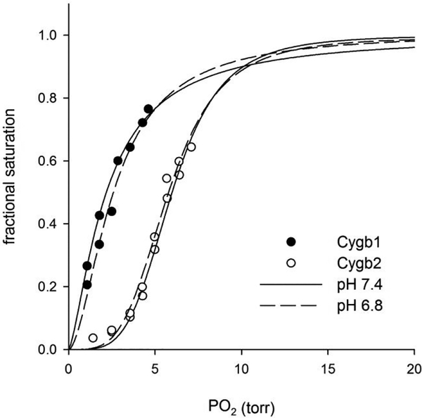
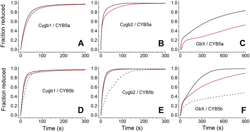
Cytoglobin is a heme protein evolutionarily related to hemoglobin and myoglobin. Cytoglobin is expressed ubiquitously in mammalian tissues; however, its physiological functions are yet unclear. Phylogenetic analyses indicate that the cytoglobin gene is highly conserved in vertebrate clades, from fish to reptiles, amphibians, birds, and mammals. Most proposed roles for cytoglobin require the maintenance of a pool of reduced cytoglobin (FeII). We have shown previously that the human cytochrome b5/cytochrome b5 reductase system, considered a quintessential hemoglobin/myoglobin reductant, can reduce human and zebrafish cytoglobins ≤250-fold faster than human hemoglobin or myoglobin. It was unclear whether this reduction of zebrafish cytoglobins by mammalian proteins indicates a conserved pathway through vertebrate evolution. Here, we report the reduction of zebrafish cytoglobins 1 and 2 by the zebrafish cytochrome b5 reductase and the two zebrafish cytochrome b5 isoforms. In addition, the reducing system also supports reduction of Globin X, a conserved globin in fish and amphibians. Indeed, the zebrafish reducing system can maintain a fully reduced pool for both cytoglobins, and both cytochrome b5 isoforms can support this process. We determined the P50 for oxygen to be 0.5 Torr for cytoglobin 1 and 4.4 Torr for cytoglobin 2 at 25 °C. Thus, even at low oxygen tensions, the reduced cytoglobins may exist in a predominant oxygen-bound form. Under these conditions, the cytochrome b5/cytochrome b5 reductase system can support a conserved role for cytoglobins through evolution, providing electrons for redox signaling reactions such as nitric oxide dioxygenation, nitrite reduction, and phospholipid oxidation.


Conflict of interest statement

CONFLICT OF INTEREST STATEMENT

A.W.D., J.J.R., M.T.G. and J.T. are coinventors of provisional and pending patents for the use of recombinant cytoglobin, neuroglobin and other heme-based molecules as antidotes for carbon monoxide poisoning. Globin Solutions, Inc. has licensed this technology. J.J.R., M.T.G. and J.T. are shareholders and officers in Globin Solutions, Inc. J.J.R. is an officer and director of Globin Solutions, Inc. M.T.G. is a director and advisor of Globin Solutions, Inc. J.T. is an officer of Globin Solutions, Inc. The remaining authors have disclosed that they do not have any potential conflicts of interest.

Figures

Figure 1.
Figure 1.. Alignment of cytochrome b5 reductase and cytochromes b5a and b5b protein sequences for zebrafish, human, and rat.
A; alignment of CYB5R3 sequences (Uniprot accession codes: zebrafish, Q6NYE6; human P00387; rat, P20070). Initial portions in italics indicate the missing residues in the soluble isoform (CYB5R3–2) of the mammalian proteins and the homologous residues in zebrafish protein; the first amino acid in the recombinant protein used in this work and the initial amino acid in the mammalian isoform 2 are marked in bold and yellow background. B; alignment of CYB5a (microsomal CYB5) and CYB5b (outer membrane CYB5) sequences (Uniprot accession codes: zebrafish b5a, Q7T341; human b5a, P00167; rat b5a, P00173; zebrafish b5b, Q6NY41; human b5b, O43169; rat b5b, P04166). Conserved heme-binding histidines are indicated in blue background. Amino acids in italics indicate sequences removed in the N-termini of CYB5b and the membrane binding C-terminal regions of CYB5a and CYB5b. The putative membrane intercalating helix region is shown in a rectangle. The first and last amino acids in the recombinant proteins used in this work, along with the splicing sites for the soluble isoform of mammalian cytochrome b5a proteins are marked in bold and yellow background; the first and last amino acids in the recombinant proteins used in other works, are indicated in bold with green background.
Figure 2.
Figure 2.. Spectral properties of zebrafish cytochrome b5 reductase and cytochromes b5a and b5b.
Panel A, CYB5R; Panel B, CYB5a; Panel C, CYB5b. The red traces represent the reduced state (FADH2 for CYB5R or ferrous (FeII) heme for CYB5a and CYB5b) and the black traces represent the oxidized proteins (FADH2 for CYB5R or ferric (FeIII) heme for CYB5a and CYB5b). Insets in Panels B and C show the 450–700nm range
Figure 3.
Figure 3.. Steady state kinetics of zebrafish Cytochrome b5 reductase.
Panels A and B, determination of the Vmax and KM towards NADH for the NADH-diaphorase activity of CYB5R with DCPIP (Panel A) or ferricyanide (Panel B) as electron acceptor. Panels C and D, determination of the Vmax and KM towards zebrafish CYB5a (Panel C) or CYB5b (Panel D) in the presence of saturating concentrations of NADH (60 μM). Experiments conducted in 50 mM Bis-Tris propane, pH 7.4, 25 °C.
Figure 4.
Figure 4.. Redox potentials of zebrafish Cytochrome b5 reductase and cytochromes b5a and b5b.
Panel A, CYB5R; Panel B, CYB5a; Panel C, CYB5b. The plots show the fit of the fraction reduced (as determined from absorbance spectra, open circles) to the Nernst equation (solid lines). The insets show the absorbance changes during the reductive titrations. Peaks at 510nm (CYB5R) or 590nm (CYB5a/b) are due to the mediators.
Figure 5.
Figure 5.. Thermal denaturation of zebrafish Cytochrome b5a and cytochrome b5b.
Panel A, CYB5a; Panel B, CYB5b. The spectra obtained at increasing temperatures (20–95 °C) are shown, the direction of the absorbance changes is indicated by arrows. The insets show the changes in the Soret peak absorbance with the temperature and the fit of the absorbance changes to the Santoro-Bolen equation (red lines).
Figure 6.
Figure 6.. Oxygen equilibrium curves of zebrafish cytoglobins 1 and 2 at 37 °C.
The points for Cygb1 (solid circles) and Cygb2 (open circles) are indicated. The lines show the fit to the Hill sigmoidal equation.
Figure 7.
Figure 7.. Reduction of zebrafish cytoglobins 1 and 2 and GlobinX by cytochrome b5 reductase and cytochrome b5.
Panel A, Reduction of Cygb1 by zebrafish CYB5a and CYB5R. Panel B, Reduction of Cygb2 by zebrafish CYB5a and CYB5R. Panel C, Reduction of GlobinX by zebrafish CYB5a and CYB5R, Panel D, Reduction of Cygb1 by zebrafish CYB5b and CYB5R (solid lines) or the human CYB5b/CYB5R system (dotted lines). Panel E, Reduction of Cygb2 by zebrafish CYB5b and CYB5R (solid lines) or the human CYB5b/CYB5R system (dotted lines). Panel F, Reduction of GlobinX by zebrafish CYB5b and CYB5R (solid lines) or the human CYB5b/CYB5R system (dotted lines). Red lines, reactions monitored at 25 °C, Black lines, reactions monitored at 37 °C.

References

    1. Burmester T, and Hankeln T (2014) Function and evolution of vertebrate globins, Acta Physiol (Oxf) 211, 501–514. - PubMed
    1. Hoogewijs D, Ebner B, Germani F, Hoffmann FG, Fabrizius A, Moens L, Burmester T, Dewilde S, Storz JF, Vinogradov SN, and Hankeln T (2012) Androglobin: a chimeric globin in metazoans that is preferentially expressed in Mammalian testes, Mol Biol Evol 29, 1105–1114. - PMC - PubMed
    1. Gardner AM, Cook MR, and Gardner PR (2010) Nitric-oxide dioxygenase function of human cytoglobin with cellular reductants and in rat hepatocytes, J Biol Chem 285, 23850–23857. - PMC - PubMed
    1. Reeder BJ, Svistunenko DA, and Wilson MT (2011) Lipid binding to cytoglobin leads to a change in haem co-ordination: a role for cytoglobin in lipid signalling of oxidative stress, Biochem J 434, 483–492. - PubMed
    1. Tejero J, Kapralov AA, Baumgartner MP, Sparacino-Watkins CE, Anthonymuthu TS, Vlasova II, Camacho CJ, Gladwin MT, Bayir H, and Kagan VE (2016) Peroxidase Activation of Cytoglobin by Anionic phospholipids: Mechanisms and Consequences, Biochimica et Biophysica Acta-Molecular and Cell Biology of Lipids 1861, 391–401. - PMC - PubMed

Publication types