Review
. 2019 Jun 14:13:600.
doi: 10.3389/fnins.2019.00600. eCollection 2019.

Mechanics of Brain Tissues Studied by Atomic Force Microscopy: A Perspective

Affiliations
Review

Mechanics of Brain Tissues Studied by Atomic Force Microscopy: A Perspective

Prem Kumar Viji Babu et al. Front Neurosci. .

Abstract

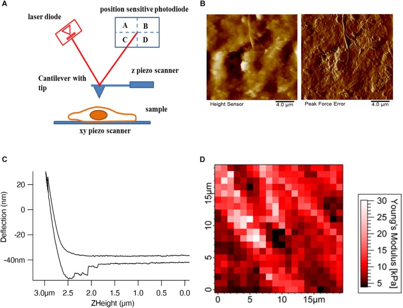
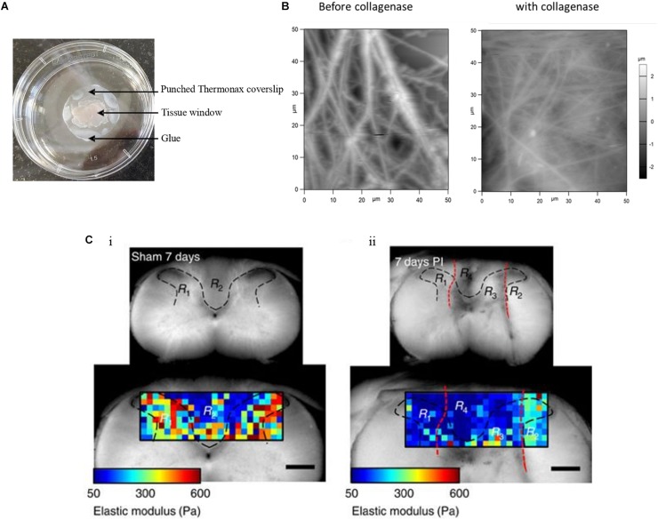
Tissue morphology and mechanics are crucial to the regulation of organ function. Investigating the exceptionally complex tissue of the brain at the sub-micron scale is challenging due to the complex structure and softness of this tissue, despite the large interest of biologists, medical engineers, biophysicists, and others in this topic. Atomic force microscopy (AFM) both as an imaging and as a mechanical tool provides an excellent opportunity to study soft biological samples such as live brain tissues. Here we review the principles of AFM, the performance of AFM in tissue imaging and mechanical mapping of cells and tissues, and finally opening the prospects and challenges of probing the biophysical properties of brain tissue using AFM.

Keywords: atomic force microscopy (AFM); mechanical mapping; tissue imaging; tissue mechanics; tissue morphology.


Figures

FIGURE 1
FIGURE 1
AFM setup and tissue imaging. (A) AFM consists of four important components – cantilever with an AFM tip, laser diode, position-sensitive photodetector, and xyz-piezo scanner. (B) Height and deflection images show the decellularized dermal matrix which shows the collagen fibers running along other matrix components (Rianna et al., 2018). (C) Deflection (nm) vs. Z height (μm) shows the approach and retract curve (Viji Babu et al., 2018). (D) Force maps show the elastic modulus values of mouse skin tissue in a respective color scale (Pa) (Joshi et al., 2017).
FIGURE 2
FIGURE 2
Tissue sample immobilization and AFM imaging of mouse skin tissue sample. (A) Tissue samples are immobilized with punched Thermanox coverslips, glued to the Petri dish at their borders, thus avoiding direct contact of tissue with glue. AFM tip accesses the sample through the tissue window. (B) AFM height images show the presence of thick and rich ECM fibers in the mouse skin tissue matrix before addition of collagenase and after the addition of thick fibers, after which disappeared and decreased ECM fibers are seen (Joshi et al., 2017). (C) The gray (R1 and R3 in both i and ii) and white (R2 and R4 in both i and ii) matter are indicated within the area (black dashed lines) of the transverse spinal cord section of a sham control and of an animal with a dorsal column crush lesion at 7 days post-injury. The elastic moduli values are represented by means of a color map. In ii, the injured areas are identified by the red dashed lines (Reprinted with the permission from Moeendarbary et al., 2017 and this work is licensed under a Creative Commons Attribution 4.0 International License. To view the copy of this license, visit http://creativecommons.org/licenses/by/4.0/).

Similar articles

Cited by

References

    1. Ando T. (2018). High-speed atomic force microscopy and its future prospects. Biophys. Rev. 10 285–292. 10.1007/s12551-017-0356-355 - DOI - PMC - PubMed
    1. Benoit M., Gabriel D., Gerisch G., Gaub H. E. (2000). Discrete interactions in cell adhesion measured by single-molecule force spectroscopy. Nat. Cell Biol. 2 313–317. 10.1038/35014000 - DOI - PubMed
    1. Bouchonville N., Meyer M., Gaude C., Gay E., Ratel D., Nicolas A. (2016). AFM mapping of the elastic properties of brain tissue reveals kPa μm-1gradients of rigidity. Soft Matter 12 6232–6239. 10.1039/c6sm00582a - DOI - PubMed
    1. Braet F., Rotsch C., Wisse E., Radmacher M. (1998). Comparison of fixed and living liver endothelial cells by atomic force microscopy. Appl. Phys. A 66 S575–S578. 10.1007/s003390051204 - DOI
    1. Chan S., Neu C., DuRaine G., Komvopoulos K., Reddi A. (2010). Atomic force microscope investigation of the boundary-lubricant layer in articular cartilage. Osteoarthritis Cartilage 18 956–963. 10.1016/j.joca.2010.03.012 - DOI - PubMed