

Identification of Autoreactive B Cell Subpopulations in Peripheral Blood of Autoimmune Patients With Pemphigus Vulgaris
- PMID: 31258541
- PMCID: PMC6587433
- DOI: 10.3389/fimmu.2019.01375
Identification of Autoreactive B Cell Subpopulations in Peripheral Blood of Autoimmune Patients With Pemphigus Vulgaris
Abstract
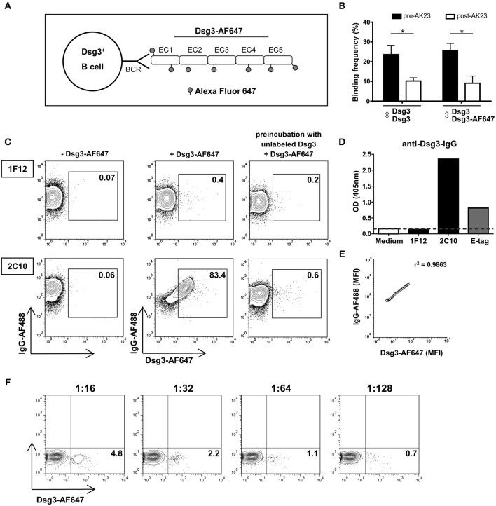
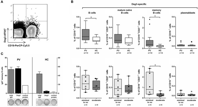
Pemphigus vulgaris (PV) is a rare blistering disease caused by IgG autoantibodies against the epidermal adhesion molecules desmoglein (Dsg)3 and Dsg1 providing a well-characterized paradigm of an antibody-mediated organ-specific autoimmune disease. In PV patients who have achieved clinical remission after B cell-depleting therapy, relapses often coincide with a reoccurrence of B cells and Dsg-specific autoantibodies. Here, we analyzed Dsg3-specific B cell subpopulations (i.e., total CD19+ B cells, CD19+CD27-B cells, CD19+CD27+ memory B cells, and CD19+CD27hiCD38hi plasmablasts) in peripheral blood of both PV patients (n = 14) at different stages of disease and healthy individuals (n = 14) by flow cytometry using fluorescently labeled recombinant human Dsg3 protein. Applying this approach, Dsg3-specific B cells could be detected at low frequencies (0.11-0.53% of CD19+ B cells) and numbers of Dsg3-specific memory B cells were significantly increased in PV patients in clinical remission receiving minimal immunosuppressive therapy. Finally, we confirmed in vitro that Dsg3-reactive memory B cells were able to produce anti-Dsg3 IgG autoantibodies upon ex vivo activation. Thus, monitoring of Dsg3-specific B cells in PV is of particular interest to further characterize the immunopathogenesis of PV.
Keywords: B cells; autoimmunity; desmoglein 3; flow cytometry; pemphigus vulgaris.
Figures
Similar articles
-
Pathogenic IgG antibodies against desmoglein 3 in pemphigus vulgaris are regulated by HLA-DRB1*04:02-restricted T cells.J Immunol. 2014 Nov 1;193(9):4391-9. doi: 10.4049/jimmunol.1401081. Epub 2014 Sep 24. J Immunol. 2014. PMID: 25252957
-
Immunophenotyping in pemphigus reveals a TH17/TFH17 cell-dominated immune response promoting desmoglein1/3-specific autoantibody production.J Allergy Clin Immunol. 2021 Jun;147(6):2358-2369. doi: 10.1016/j.jaci.2020.11.008. Epub 2020 Nov 20. J Allergy Clin Immunol. 2021. PMID: 33221382
-
[Detection of serum desmoglein antibody level using enzyme-linked immunosorbent assay (ELISA) for monitoring disease activity in patients with pemphigus vulgaris].Beijing Da Xue Xue Bao Yi Xue Ban. 2011 Jun 18;43(3):414-5. Beijing Da Xue Xue Bao Yi Xue Ban. 2011. PMID: 21681274 Chinese.
-
Pemphigus: a Comprehensive Review on Pathogenesis, Clinical Presentation and Novel Therapeutic Approaches.Clin Rev Allergy Immunol. 2018 Feb;54(1):1-25. doi: 10.1007/s12016-017-8662-z. Clin Rev Allergy Immunol. 2018. PMID: 29313220 Review.
-
Analysis of the T cells that are potentially involved in autoantibody production in pemphigus vulgaris.J Dermatol. 1999 Nov;26(11):748-52. doi: 10.1111/j.1346-8138.1999.tb02086.x. J Dermatol. 1999. PMID: 10635617 Review.
Cited by
-
From neglect to spotlight: the underappreciated role of B cells in cutaneous inflammatory diseases.Front Immunol. 2024 Feb 15;15:1328785. doi: 10.3389/fimmu.2024.1328785. eCollection 2024. Front Immunol. 2024. PMID: 38426103 Free PMC article. Review.
-
Efgartigimod as a novel FcRn inhibitor for autoimmune disease.Neurol Sci. 2024 Sep;45(9):4229-4241. doi: 10.1007/s10072-024-07460-5. Epub 2024 Apr 22. Neurol Sci. 2024. PMID: 38644454 Review.
-
Skin-Associated B Cells in the Pathogenesis of Cutaneous Autoimmune Diseases-Implications for Therapeutic Approaches.Cells. 2020 Dec 7;9(12):2627. doi: 10.3390/cells9122627. Cells. 2020. PMID: 33297481 Free PMC article. Review.
-
Engaging T cells for cleanup.Front Immunol. 2025 May 6;16:1551424. doi: 10.3389/fimmu.2025.1551424. eCollection 2025. Front Immunol. 2025. PMID: 40416957 Free PMC article. Review.
-
FcRn Antagonism Leads to a Decrease of Desmoglein-Specific B Cells: Secondary Analysis of a Phase 2 Study of Efgartigimod in Pemphigus Vulgaris and Pemphigus Foliaceus.Front Immunol. 2022 May 18;13:863095. doi: 10.3389/fimmu.2022.863095. eCollection 2022. Front Immunol. 2022. PMID: 35663943 Free PMC article. Clinical Trial.
References
Publication types
MeSH terms
Substances
LinkOut - more resources
Full Text Sources
Other Literature Sources
Medical
Research Materials
Miscellaneous