

Altered spontaneous brain activity patterns in patients with corneal ulcer using amplitude of low-frequency fluctuation: An fMRI study
- PMID: 31258645
- PMCID: PMC6566102
- DOI: 10.3892/etm.2019.7550
Altered spontaneous brain activity patterns in patients with corneal ulcer using amplitude of low-frequency fluctuation: An fMRI study
Abstract
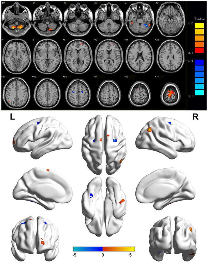
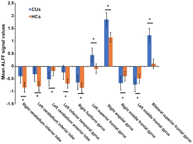
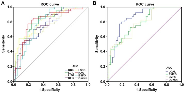
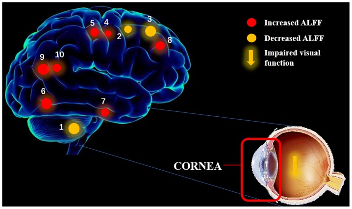
The aim of the present study was to investigate the altered spontaneous brain activity in patients with corneal ulcer (CU) through the amplitude of low-frequency fluctuation (ALFF) technique and the association with their visual performance. A total of 40 patients with CU and 40 healthy controls (HCs) matched for sex, age and educational level were enrolled. Resting-state functional magnetic resonance imaging (rs-fMRI) was performed to examine the probands. Spontaneous cerebral activity variations were investigated using the ALFF technique. The average ALFF values of the CU patients and the HCs were classified by utilizing receiver operating characteristic (ROC) curves. Contrary to HCs, the CU patients had significantly lower ALFF values in the left cerebellar anterior lobe, right middle frontal gyrus and left middle frontal gyrus, but higher ALFF values in the right cerebellar inferior lobe, left cerebellar inferior lobe, left inferior temporal gyrus, right fusiform gyrus, left superior frontal gyrus, right angular gyrus and bilateral superior frontal gyrus. ROC curve analysis of each brain region indicated that the accuracy of ALFF value specificity between the CU and HCs of the area under the curve was perfect. In conclusion, abnormal spontaneous activities were detected in numerous brain regions of CU patients, which may provide useful information for understanding the dysfunction of CU. These activity changes in brain regions may be used as effective clinical indicators for CU.
Keywords: ALFF; corneal ulcer; fMRI; resting state; spontaneous activity.
Figures
Similar articles
-
Altered spontaneous brain activity patterns in patients with retinal vein occlusion indicated by the amplitude of low-frequency fluctuation: A functional magnetic resonance imaging study.Exp Ther Med. 2019 Sep;18(3):2063-2071. doi: 10.3892/etm.2019.7770. Epub 2019 Jul 12. Exp Ther Med. 2019. PMID: 31410162 Free PMC article.
-
The predictive potential of altered spontaneous brain activity patterns in diabetic retinopathy and nephropathy.EPMA J. 2019 Jul 5;10(3):249-259. doi: 10.1007/s13167-019-00171-4. eCollection 2019 Sep. EPMA J. 2019. PMID: 31462942 Free PMC article.
-
Disturbed spontaneous brain-activity pattern in patients with optic neuritis using amplitude of low-frequency fluctuation: a functional magnetic resonance imaging study.Neuropsychiatr Dis Treat. 2015 Dec 16;11:3075-83. doi: 10.2147/NDT.S92497. eCollection 2015. Neuropsychiatr Dis Treat. 2015. PMID: 26719692 Free PMC article.
-
Altered spontaneous brain activity in lumbar disc herniation patients: insights from an ALE meta-analysis of neuroimaging data.Front Neurosci. 2024 Feb 6;18:1349512. doi: 10.3389/fnins.2024.1349512. eCollection 2024. Front Neurosci. 2024. PMID: 38379762 Free PMC article.
-
Commonalities and distinctions between the type 2 diabetes mellitus and Alzheimer's disease: a systematic review and multimodal neuroimaging meta-analysis.Front Neurosci. 2023 Dec 6;17:1301778. doi: 10.3389/fnins.2023.1301778. eCollection 2023. Front Neurosci. 2023. PMID: 38125399 Free PMC article. Review.
Cited by
-
Alternation of brain intrinsic activity in patients with hypertensive retinopathy: a resting-state fMRI study.Aging (Albany NY). 2021 Sep 13;13(17):21659-21670. doi: 10.18632/aging.203510. Epub 2021 Sep 13. Aging (Albany NY). 2021. PMID: 34516404 Free PMC article.
-
Altered spontaneous brain activity patterns in diabetic patients with vitreous hemorrhage using amplitude of low‑frequency fluctuation: A resting‑state fMRI study.Mol Med Rep. 2020 Sep;22(3):2291-2299. doi: 10.3892/mmr.2020.11294. Epub 2020 Jul 3. Mol Med Rep. 2020. PMID: 32705185 Free PMC article.
-
Brain Activity Changes in Slow 5 and Slow 4 Frequencies in Patients With Optic Neuritis: A Resting State Functional MRI Study.Front Neurol. 2022 Feb 21;13:823919. doi: 10.3389/fneur.2022.823919. eCollection 2022. Front Neurol. 2022. PMID: 35265028 Free PMC article.
-
Children with strabismus and amblyopia presented abnormal spontaneous brain activities measured through fractional amplitude of low-frequency fluctuation (fALFF).Front Neurol. 2022 Aug 12;13:967794. doi: 10.3389/fneur.2022.967794. eCollection 2022. Front Neurol. 2022. PMID: 36034279 Free PMC article.
-
Amplitude of low-frequency fluctuation-based regional radiomics similarity network: Biomarker for Parkinson's disease.Heliyon. 2023 Mar 6;9(3):e14325. doi: 10.1016/j.heliyon.2023.e14325. eCollection 2023 Mar. Heliyon. 2023. PMID: 36950566 Free PMC article.
References
-
- World Health Organization, corp-author. Causes of blindness and visual impairment. 2016
-
- El Sheha H. Self-retained amniotic membrane for dendritic keratitis. Ascrs. 2015
-
- Mascarenhas J, Lalitha P, Prajna NV, Srinivasan M, Das M, D'Silva SS, Oldenburg CE, Borkar DS, Esterberg EJ, Lietman TM, Keenan JD. Acanthamoeba, fungal, and bacterial keratitis: A comparison of risk factors and clinical features. Am J Ophthalmol. 2014;157:56–62. doi: 10.1016/j.ajo.2013.08.032. - DOI - PMC - PubMed
LinkOut - more resources
Full Text Sources