

Photothermal effect by 808-nm laser irradiation of melanin: a proof-of-concept study of photothermal therapy using B16-F10 melanotic melanoma growing in BALB/c mice
- PMID: 31259063
- PMCID: PMC6583352
- DOI: 10.1364/BOE.10.002932
Photothermal effect by 808-nm laser irradiation of melanin: a proof-of-concept study of photothermal therapy using B16-F10 melanotic melanoma growing in BALB/c mice
Abstract
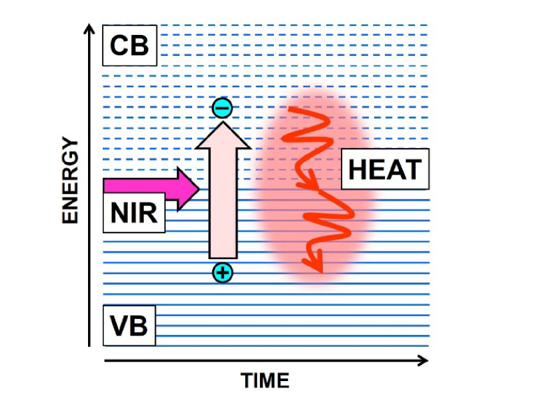
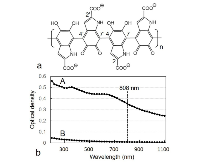
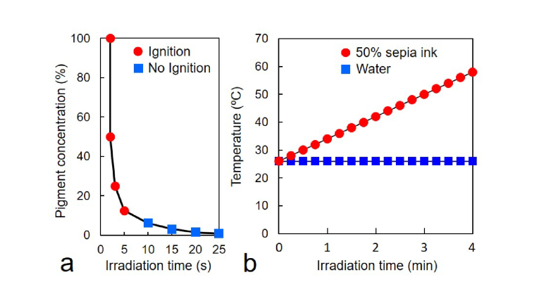
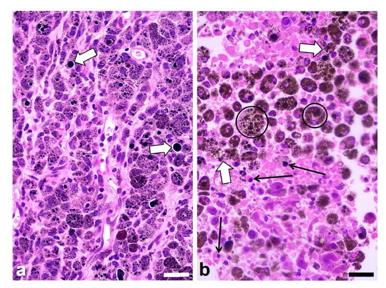
The photothermal effect is undergoing great interest due to advances in new photosensitizing materials and better-suited light sources, but studies are frequently hampered by the need to employ exogenous photothermal agents and expensive irradiation devices. Here we present a simple strategy based on direct NIR irradiation of the melanin pigment with a commercial 808-nm laser pointer. Proof-of-concept studies showed efficient photothermal effects on melanin in vitro and in vivo. After NIR irradiation, BALB/c mice bearing B16-F10 melanotic melanoma tumors revealed severe histopathological damage and massive necrosis in melanin-containing tumor tissue, while surrounding healthy tissues showed no damage. Therefore, the feasibility of this approach may allow implementing direct procedures for photothermal therapy of pigmented tumors.
Conflict of interest statement
The authors declare that there are no conflicts of interest related to this article.
Figures
Similar articles
-
NIR laser pointer for in vivo photothermal therapy of murine LM3 tumor using intratumoral China ink as a photothermal agent.Lasers Med Sci. 2018 Aug;33(6):1307-1315. doi: 10.1007/s10103-018-2483-z. Epub 2018 Mar 16. Lasers Med Sci. 2018. PMID: 29549555
-
Laser-induced vapor nanobubbles for B16-F10 melanoma cell killing and intracellular delivery of chemotherapeutics.J Control Release. 2024 Jan;365:1019-1036. doi: 10.1016/j.jconrel.2023.12.006. Epub 2023 Dec 22. J Control Release. 2024. PMID: 38065413
-
Utilizing 808 nm laser for sensitizing of melanoma tumors to megavoltage radiation therapy.Lasers Med Sci. 2020 Feb;35(1):87-93. doi: 10.1007/s10103-019-02796-3. Epub 2019 May 10. Lasers Med Sci. 2020. PMID: 31076924
-
In vitro and in vivo tumor annihilation by near-infrared photothermal effect of a NiFe2O4/C nanocomposite.Colloids Surf B Biointerfaces. 2018 Oct 1;170:393-400. doi: 10.1016/j.colsurfb.2018.06.034. Epub 2018 Jun 19. Colloids Surf B Biointerfaces. 2018. PMID: 29945051
-
Evaluation of a nanocomposite of PEG-curcumin-gold nanoparticles as a near-infrared photothermal agent: an in vitro and animal model investigation.Lasers Med Sci. 2018 Nov;33(8):1769-1779. doi: 10.1007/s10103-018-2538-1. Epub 2018 May 22. Lasers Med Sci. 2018. PMID: 29790012
Cited by
-
Chromophore-Targeting Precision Antimicrobial Phototherapy.Cells. 2023 Nov 20;12(22):2664. doi: 10.3390/cells12222664. Cells. 2023. PMID: 37998399 Free PMC article. Review.
-
Photothermal treatment-based heat stress regulates function of myeloid-derived suppressor cells.Sci Rep. 2024 Aug 14;14(1):18847. doi: 10.1038/s41598-024-69074-3. Sci Rep. 2024. PMID: 39143087 Free PMC article.
-
Sema4D silencing increases the sensitivity of nivolumab to B16-F10 resistant melanoma via inhibiting the PI3K/AKT signaling pathway.PeerJ. 2023 Apr 19;11:e15172. doi: 10.7717/peerj.15172. eCollection 2023. PeerJ. 2023. PMID: 37096066 Free PMC article.
-
Photothermal immunotherapy of melanoma using TLR-7 agonist laden tobacco mosaic virus with polydopamine coat.Nanomedicine. 2022 Aug;44:102573. doi: 10.1016/j.nano.2022.102573. Epub 2022 Jun 18. Nanomedicine. 2022. PMID: 35728739 Free PMC article.
-
Bio-Applications of Multifunctional Melanin Nanoparticles: From Nanomedicine to Nanocosmetics.Nanomaterials (Basel). 2020 Nov 17;10(11):2276. doi: 10.3390/nano10112276. Nanomaterials (Basel). 2020. PMID: 33212974 Free PMC article. Review.
References
-
- Guy G. P., Jr., Thomas C. C., Thompson T., Watson M., Massetti G. M., Richardson L. C., Centers for Disease Control and Prevention (CDC) , “Vital signs: melanoma incidence and mortality trends and projections - United States, 1982-2030,” MMWR Morb. Mortal. Wkly. Rep. 64(21), 591–596 (2015). - PMC - PubMed
-
- Kabigting F. D., Nelson F. P., Kauffman C. L., Popoveniuc G., Dasanu C. A., Alexandrescu D. T., “Malignant melanoma in African-Americans,” Dermatol. Online J. 15(2), 3 (2009). - PubMed
LinkOut - more resources
Full Text Sources
Miscellaneous