An official website of the United States government
The .gov means it’s official.
Federal government websites often end in .gov or .mil. Before
sharing sensitive information, make sure you’re on a federal
government site.
The site is secure.
The https:// ensures that you are connecting to the
official website and that any information you provide is encrypted
and transmitted securely.
We report the case of a 9-month-old male with Williams syndrome who underwent patch augmentation of supravalvar aortic stenosis and pulmonary artery stenosis, and required emergent drug-eluting left coronary artery stenting on post-operative day 1 for severe left ventricular dysfunction related to myocardial ischemia.
Keywords:
coronary artery disease; percutaneous coronary intervention; williams syndrome.
Conflict of Interest: The authors have no financial conflicts of interest to disclose related to this case report.
Figures
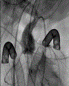
Figure 1:
Straight A/P angiography demonstrating a…
Figure 1:
Straight A/P angiography demonstrating a severely stenotic left main coronary artery (LMCA), with…
Figure 1:
Straight A/P angiography demonstrating a severely stenotic left main coronary artery (LMCA), with a proximal diameter of less than 1 mm. The white arrow denotes the area of stenosis.
Figure 2:
Straight AP angiography post-stent placement…
Figure 2:
Straight AP angiography post-stent placement and dilation demonstrates a widely patent left main…
Figure 2:
Straight AP angiography post-stent placement and dilation demonstrates a widely patent left main coronary artery without extravasation of dye, thrombus, or evidence of endothelial injury. The white arrow denotes the patent area of stented coronary artery.
Figure 3:
Angiography 7 months post procedure…
Figure 3:
Angiography 7 months post procedure with AP camera angulated RAO 30 degrees. The…
Figure 3:
Angiography 7 months post procedure with AP camera angulated RAO 30 degrees. The left main coronary remains patent without evidence of intimal stenosis, and no significant collateralization to distal left coronary system. The white arrow denotes the patent area of stented coronary artery.
Figure 4:
Parasternal short axis view demonstrating…
Figure 4:
Parasternal short axis view demonstrating stable stent placement and color Doppler flow in…
Figure 4:
Parasternal short axis view demonstrating stable stent placement and color Doppler flow in diastole, indicating stent patency. The white arrow denotes the patent left main coronary artery.
Upadhyay S, Seiden H, Epstein S, (2011) Left Main Coronary Artery Stenting After Cardiac Arrest in an Infant with Williams Syndrome. Catheter Cardiovasc Interv 78:940–944. DOI: 10.1002/ccd.23040.
-
DOI
-
PubMed
Hallbergson A, Rome J, (2015) Percutaneous Left Main Coronary Artery Stent for Acute Myocardial Ischemia after Repaired ALCAPA. Catheter Cardiovasc Interv 85:1017–1020. DOI: 10.1002/ccd.25771.
-
DOI
-
PubMed
Drossner DM, Chappell C, Rab T, Kim D (2012) Percutaneous Coronary Intervention for Acute Myocardial Infarction in a Pediatric Patient With Coronary Aneurysm and Stenosis Due to Kawasaki Disease. Pediatr Cardiol 33: 811–813. DOI: 10.1007/s00246-012-0190-2
-
DOI
-
PubMed
El-Said HG, Ebrahim M, Moore JW (2015) Left Main Coronary Artery Stenting in a 3.6 kg Infant After Arterial Switch Operation for Transposition of the Great Arteries. Ann Pediatr Cardiol 8(2): 143–146. DOI: 10.4103/0974-2069.157033.
-
DOI
-
PMC
-
PubMed
Chrysant GS, Balzer D, Taniuchi M (2005) Left Main Stem Coronary Artery Stenting in a 3-Month Old Child After Anomalous Left Coronary Artery from Pulmonary Artery Repair. Pediatr Cardiol 26: 734–736. DOI:10.1007/s00246-004-0758-6
-
DOI
-
PubMed