Review
. 2019 May 14;3(7):1302-1320.
doi: 10.1210/js.2019-00053. eCollection 2019 Jul 1.

Neuroendocrine Regulation of Brain Cytokines After Psychological Stress

Affiliations
Review

Neuroendocrine Regulation of Brain Cytokines After Psychological Stress

John D Johnson et al. J Endocr Soc. .

Abstract

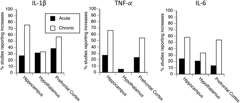
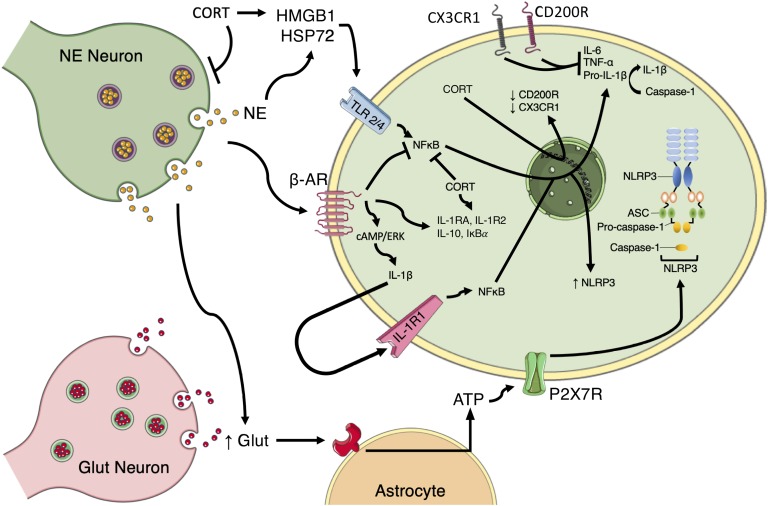
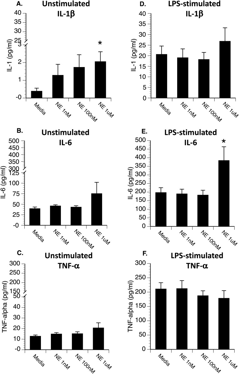
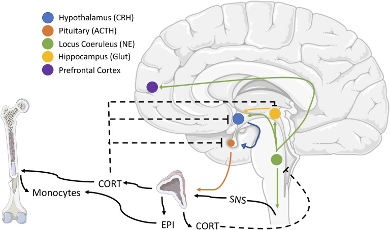
There is growing evidence that stress-induced brain cytokines are important in the etiology of depression and anxiety. Here, we review how the neuroendocrine responses to psychological stressors affect the immediate and long-term regulation of inflammatory cytokines within the brain and highlight how the regulation changes across time with repeated stress exposure. In doing so, we report on the percentage of studies in the literature that observed increases in either IL-1β, TNF-α, or IL-6 within the hypothalamus, hippocampus, or prefrontal cortex after either acute or chronic stress exposure. The key takeaway is that catecholamines and glucocorticoids play critical roles in the regulation of brain cytokines after psychological stress exposure. Central catecholamines stimulate the release of IL-1β from microglia, which is a key factor in the further activation of microglia and recruitment of monocytes into the brain. Meanwhile, the acute elevation of glucocorticoids inhibits the production of brain cytokines via two mechanisms: the suppression of noradrenergic locus coeruleus neurons and inhibition of the NFκB signaling pathway. However, glucocorticoids and peripheral catecholamines facilitate inflammatory responses to future stimuli by stimulating monocytes to leave the bone marrow, downregulating inhibitory receptors on microglia, and priming inflammatory responses mediated by peripheral monocytes or macrophages. The activation of microglia and the elevation of peripheral glucocorticoid and catecholamine levels are both necessary during times of stress exposure for the development of psychopathologies.

Keywords: IL-1β; IL-6; TNF-α; hippocampus; hypothalamus; prefrontal cortex.


Figures

Figure 1.
Figure 1.
The percentage of studies that report a significant increase in IL-1β, TNF-α, or IL-6 levels within the hippocampus, hypothalamus, or prefrontal cortex after acute or chronic stress exposure.
Figure 2.
Figure 2.
Schematic of the proposed mechanisms by which norepinephrine (NE) and corticosterone/cortisol (CORT) regulate sterile inflammation. NE promotes inflammation by (i) facilitating glutamate (GLUT) release and production of ATP by astrocytes that then promote the oligomerization of the NLRP3 inflammasome; (ii) acting at β-ARs to induce IL-1β production through a cAMP/ERK-dependent pathway, which can lead to IL-1β acting in an autocrine fashion to induce NFκB signaling pathways and production of NLRP3, IL-6, TNF-α, and pro-IL-1β; and (iii) causing the release of HSP72, which acts at TLR 2/4 to stimulate NFκB signaling. CORT promotes inflammation by (i) causing the release of HMGB1, which activates TLR 2/4 signaling pathways and (ii) downregulating CD200R and CX3CR1, which are negative regulators of inflammatory responses in microglia. NE and CORT also have anti-inflammatory properties that include inhibiting NFκB signaling and upregulating IL-1RA, IL-1R2, IL-10, and IκBα. CORT also has the ability to suppress noradrenergic activity, thereby reducing NE release. ERK, extracellular signal-regulated kinase; IL-1R2, IL-1 receptor type 2; IκBα, nuclear factor of κ light polypeptide gene enhancer in β-cells inhibitor α; CD200R, CD200 receptor; CX3CR1, CX3C receptor type 1.
Figure 3.
Figure 3.
Hippocampal microglia were plated in a 96-well culture plate at 10,000 cells per well and incubated with either (A–C) media (unstimulated) or (D–F) 0.5 μg LPS (stimulated), along with different concentrations of norepinephrine. After 20 hours, supernatants were collected for measurement of IL-1β, IL-6, and TNF-α by ELISA (R&D Systems). Under unstimulated conditions (A–C), norepinephrine resulted in (A) a dose-dependent increase in IL-1β (A). Under LPS-stimulated conditions (D–F), the highest concentration of norepinephrine significantly increased (E) LPS-induced IL-6 release. *P < 0.05 difference from media-treated cells.
Figure 4.
Figure 4.
Schematic of the effects of peripheral glucocorticoids and catecholamines on peripheral monocytes and feedback on locus coeruleus neurons. Brainstem noradrenergic neurons contribute to the activation of the hypothalamus-pituitary-adrenal (HPA) axis and activation of the sympathetic nervous system (SNS), which results in the collective release of circulating corticosterone or cortisol (CORT) and epinephrine (EPI), respectively. CORT causes monocytes to leave the bone marrow, whereas EPI primes their responses to future activation. CORT provides negative feedback at multiple levels of HPA regulation and inhibits central noradrenergic release. Negative feedback is indicated by dashed lines. CRH, corticotropin-releasing hormone; Glut, glutamate.

References

    1. Ebert DD, Buntrock C, Mortier P, Auerbach R, Weisel KK, Kessler RC, Cuijpers P, Green JG, Kiekens G, Nock MK, Demyttenaere K, Bruffaerts R. Prediction of major depressive disorder onset in college students. Depress Anxiety. 2019;36(4):294–304. - PMC - PubMed
    1. Kessler RC. The effects of stressful life events on depression. Annu Rev Psychol. 1997;48(1):191–214. - PubMed
    1. Levine J, Barak Y, Chengappa KN, Rapoport A, Rebey M, Barak V. Cerebrospinal cytokine levels in patients with acute depression. Neuropsychobiology. 1999;40(4):171–176. - PubMed
    1. Köhler CA, Freitas TH, Stubbs B, Maes M, Solmi M, Veronese N, de Andrade NQ, Morris G, Fernandes BS, Brunoni AR, Herrmann N, Raison CL, Miller BJ, Lanctôt KL, Carvalho AF. Peripheral alterations in cytokine and chemokine levels after antidepressant drug treatment for major depressive disorder: systematic review and meta-analysis. Mol Neurobiol. 2018;55(5):4195–4206. - PubMed
    1. Maes M. Evidence for an immune response in major depression: a review and hypothesis. Prog Neuropsychopharmacol Biol Psychiatry. 1995;19(1):11–38. - PubMed

LinkOut - more resources