. 2019 Oct 1;76(10):1230-1239.
doi: 10.1001/jamaneurol.2019.1708.

Progressive Cortical Thinning in Patients With Focal Epilepsy

Affiliations

Progressive Cortical Thinning in Patients With Focal Epilepsy

Marian Galovic et al. JAMA Neurol. .

Abstract

Importance: It is controversial whether epilepsy is a static or progressive disease. Evidence of progressive gray matter loss in epilepsy would support early diagnosis, rapid treatment, and early referral for surgical interventions.

Objective: To demonstrate progressive cortical thinning in patients with focal epilepsy distinct from cortical thinning associated with normal aging.

Design, setting, and participants: A case-control neuroimaging study was conducted from August 3, 2004, to January 26, 2016, among 190 patients with focal epilepsy at a tertiary epilepsy referral center (epilepsy data) and 3 independent comparison cohorts matched for age and sex (healthy volunteer data; n = 141).

Exposures: Two or more high-resolution T1-weighted magnetic resonance imaging scans at least 6 months apart (mean [SD] interval, 2.5 [1.6] years).

Main outcomes and measures: Global and vertexwise rate of progressive cortical thinning.

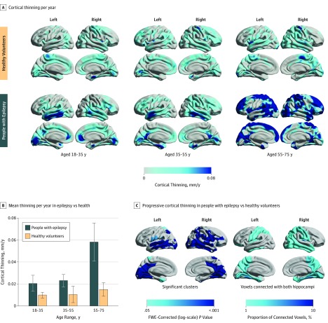
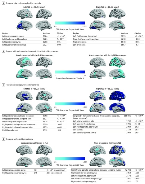
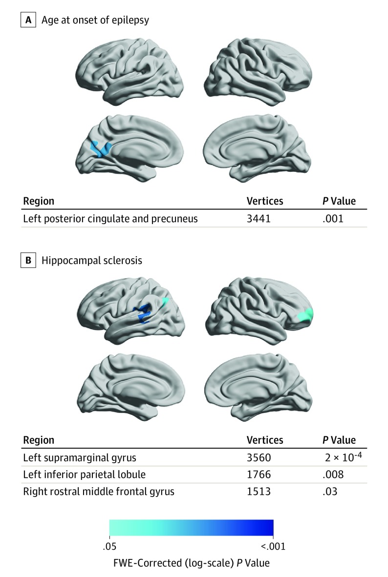
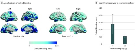
Results: A total of 190 people with focal epilepsy (99 women and 91 men; mean [SD] age, 36 [11] years; 396 magnetic resonance imaging scans) were compared with 141 healthy volunteers (76 women and 65 men; mean [SD] age, 35 [17] years; 282 magnetic resonance imaging scans). Widespread highly significant progressive cortical thinning exceeding normal aging effects, mainly involving the bilateral temporal lobes, medial parietal and occipital cortices, pericentral gyri, and opercula, was seen in 146 individuals with epilepsy (76.8%; 95% CI, 58%-95%). The mean (SD) annualized rate of global cortical thinning in patients with epilepsy was twice the rate of age-associated thinning observed in healthy volunteers (0.024 [0.061] vs 0.011 [0.029] mm/y; P = .01). Progression was most pronounced in adults older than 55 years and during the first 5 years after the onset of seizures. Areas of accelerated cortical thinning were detected in patients with early onset of epilepsy and in patients with hippocampal sclerosis. Accelerated thinning was not associated with seizure frequency, history of generalized seizures, or antiepileptic drug load and did not differ between patients with or without ongoing seizures. Progressive atrophy in temporal (n = 101) and frontal (n = 28) lobe epilepsy was most pronounced ipsilaterally to the epileptic focus but also affected a widespread area extending beyond the focus and commonly affected the contralateral hemisphere. For patients with temporal lobe epilepsy, accelerated cortical thinning was observed within areas structurally connected with the ipsilateral hippocampus.

Conclusions and relevance: Widespread progressive cortical thinning exceeding that seen with normal aging may occur in patients with focal epilepsy. These findings appear to highlight the need to develop epilepsy disease-modifying treatments to disrupt or slow ongoing atrophy. Longitudinal cortical thickness measurements may have the potential to serve as biomarkers for such studies.


Conflict of interest statement

Conflict of Interest Disclosures: Dr Galovic reported receiving grant MR/L013215/1 from the Medical Research Council (MRC) outside the submitted work. Dr Vos reported receiving grants from University College London/University College London Hospital (UCLH) National Institute for Health Research (NIHR) Biomedical Research Centres (BRC) during the conduct of the study. Dr Duncan reported receiving grants from National Institute for Health Research during the conduct of the study and grants from Wellcome Trust and MRC outside the submitted work. No other disclosures were reported.

Figures

Figure 1.
Figure 1.. Progressive Epilepsy-Associated Cortical Thinning Compared With Thinning Associated With Normal Aging
A, Annualized rate of cortical thinning in healthy volunteers (top) and people with epilepsy (bottom), stratified to 3 age groups (18 to <35, 35 to <55, and 55 to <75 years). B, Comparison of annualized cortical thinning rates between people with epilepsy and controls (vertical lines indicate SEM). C, Vertexwise statistical comparison of progressive cortical thinning in people with epilepsy vs healthy volunteers using linear mixed-effects models. Hemispheric surface templates are displaying a map of significant clusters after random field theory correction for multiple comparisons (left). Structural connectivity with left and right hippocampi in 10 healthy volunteers is presented as the regional proportion of connected voxels (right). FWE indicates familywise error.
Figure 2.
Figure 2.. Localization and Lateralization of Epilepsy and Their Association With Progressive Cortical Thinning
A, Comparison of left and right temporal lobe epilepsy vs healthy controls. B, Structural connectivity with the left and right hippocampi in 10 healthy volunteers is presented as the regional proportion of connected voxels. C, Comparison of left and right frontal lobe epilepsy vs healthy controls. D, Comparison of temporal vs frontal lobe epilepsy. Significant clusters (P < .05; random field theory corrected) are displayed on hemispheric surface templates and in overview tables. FLE indicates frontal lobe epilepsy; FWE, familywise error; and TLE, temporal lobe epilepsy.
Figure 3.
Figure 3.. Clinical Characteristics in People With Epilepsy and Their Association With Progressive Cortical Thinning
A, Association of progressive cortical thinning in people with epilepsy (n = 190) with younger age at onset of epilepsy. B, Association of progressive cortical thinning in people with epilepsy with presence of hippocampal sclerosis. Significant clusters (P < .05; random field theory corrected) are displayed on hemispheric surface templates and in overview tables. No areas of progressive thinning associated with seizure frequency, history of secondary generalized seizures, and number of antiepileptic drugs taken were detected, and there were no differences between patients with vs without ongoing seizures. FWE indicates familywise error.
Figure 4.
Figure 4.. Duration of Epilepsy and Its Association With Progressive Cortical Thinning
A, Annualized rate of cortical thinning in people with epilepsy with disease duration of less than 5 years or 5 years or more after the onset of first seizure. B, Comparison of annualized cortical thinning rates in people with epilepsy with short and long disease duration (vertical lines indicate SEM).

References

    1. Gowers WR. Epilepsy and Other Chronic Convulsive Disorders. London, UK: J. & A. Churchill; 1881.
    1. Rossini L, Garbelli R, Gnatkovsky V, et al. . Seizure activity per se does not induce tissue damage markers in human neocortical focal epilepsy. Ann Neurol. 2017;82(3):331-341. doi:10.1002/ana.25005 - DOI - PubMed
    1. Sutula TP, Hagen J, Pitkänen A. Do epileptic seizures damage the brain? Curr Opin Neurol. 2003;16(2):189-195. doi:10.1097/00019052-200304000-00012 - DOI - PubMed
    1. Cole AJ. Is epilepsy a progressive disease? the neurobiological consequences of epilepsy. Epilepsia. 2000;41(suppl 2):S13-S22. doi:10.1111/j.1528-1157.2000.tb01520.x - DOI - PubMed
    1. Haneef Z, Stern J, Dewar S, Engel J Jr. Referral pattern for epilepsy surgery after evidence-based recommendations: a retrospective study. Neurology. 2010;75(8):699-704. doi:10.1212/WNL.0b013e3181eee457 - DOI - PMC - PubMed