Review
. 2019 Nov 15;14(11):2335-2348.
doi: 10.1021/acschembio.9b00338. Epub 2019 Jul 16.

Procaspase-3 Overexpression in Cancer: A Paradoxical Observation with Therapeutic Potential

Affiliations
Review

Procaspase-3 Overexpression in Cancer: A Paradoxical Observation with Therapeutic Potential

Matthew W Boudreau et al. ACS Chem Biol. .

Abstract

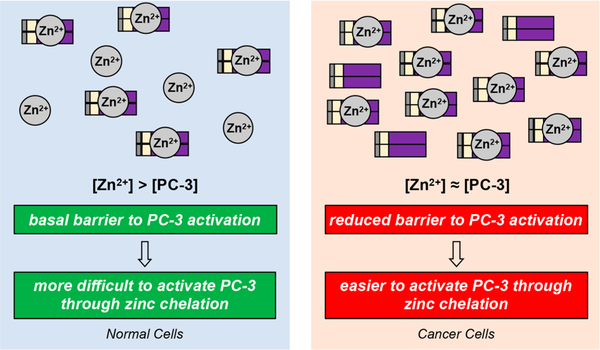
Many anticancer strategies rely on the promotion of apoptosis in cancer cells as a means to shrink tumors. Crucial for apoptotic function are executioner caspases, most notably caspase-3, that proteolyze a variety of proteins, inducing cell death. Paradoxically, overexpression of procaspase-3 (PC-3), the low-activity zymogen precursor to caspase-3, has been reported in a variety of cancer types. Until recently, this counterintuitive overexpression of a pro-apoptotic protein in cancer has been puzzling. Recent studies suggest subapoptotic caspase-3 activity may promote oncogenic transformation, a possible explanation for the enigmatic overexpression of PC-3. Herein, the overexpression of PC-3 in cancer and its mechanistic basis is reviewed; collectively, the data suggest the potential for exploitation of PC-3 overexpression with PC-3 activators as a targeted anticancer strategy.


Conflict of interest statement

The authors declare the following competing financial interest(s): The University of Illinois has filed patents on some of the technology described in this manuscript.

Figures

Figure 1.
Figure 1.
Diverse upstream mechanisms employed by cancers to prevent apoptosis. Increased expression of antiapoptotic proteins (green arrows, e.g. Bcl-2, decoy death receptors) or decreased expression or mutation of proapoptotic proteins (red arrows, x’s, e.g. APAF-1, BH3 proteins, p53) drive the net effect of preventing downstream activation of PC-3 to caspase-3 and avoiding apoptotic cell death. Protein structures displayed: p53 (PDB: 4QOl), APAF-1 (PDB: 1CY5), BH3-proteins (Bax, PDB: 1F16), decoy death receptor (Death Receptor 4, PDB: 5CIR, no reported decoy death receptor crystal structures), and Bcl-2 (PDB: 1G5M). For comprehensive reviews on the upstream signaling that results in apoptosis, see refs 14 and .
Figure 2.
Figure 2.
Autocatalytic PC-3 activation, resulting in large effects from minor initial activity. (A) Pictorial representation of PC-3 and caspase-3 domains. (B) Reaction scheme for PC-3 activation to form caspase-3. While PC-3 is canonically cleaved by caspase-8 or caspase-9, caspase-3 can be formed by PC-3-mediated proteolysis of another PC-3 protein, or caspase-3 mediated cleavage of PC-3. (C) Initial PC-3 activation to form caspase-3 leads to a proteolytic cascade to form increased levels of caspase-3, in turn increasing proteolysis.
Figure 3.
Figure 3.
pRB/E2F pathway dysregulation leading to unrestricted CASP3 gene transcription. The pRb/E2F pathway regulation of transcription and cell cycle progression. (A) The transcriptional activity of E2F is inhibited through binding with pRb with further regulation of the pathway via pl6INK4a. (D) In the presence of growth signals, cells turn on E2F transcription via CDK4/6 activation, resulting in phosphorylation of pRB and E2F translocation to the nucleus, turning on transcription of target genes (i.e., CASP3). (C) In cancer cells, the loss of pl6INK4a, pRb, or overexpression of CDK4/6 lead to relief of E2F inhibition, resulting in unregulated transcription. For a comprehensive review of this pathway in cancer, see refs 99 and .
Figure 4.
Figure 4.
Post-translational regulation of caspase-3 and PC-3 activity via inhibitory zinc. (A) Graphical representation of inhibitory zinc on caspase-3. Protein structure displayed is PDB: 2XYG. (B) Zinc binding inhibits PC-3 autocatalysis to form caspase-3. Upon zinc liberation, PC-3 can autoactivate to form caspase-3. Adapted with permission from ref 28. Copyright 2009 Elsevier.
Figure 5.
Figure 5.
Perturbation of zinc regulation via increased PC-3 levels in cancer cells. Cancer cells overexpress PC-3, making regulation of PC-3 activity through inhibitory zinc less effective. Given the autocatalytic nature of PC-3 autoactivation and amplification, these changes in zinc/PC-3 ratio may prove sufficient to alter basal PC-3 (and ultimately caspase-3) activity within a cell. Zinc (Zn2+) circles represent the labile zinc pool.
Figure 6.
Figure 6.
Promotion of DNA damage and genomic instability by subapoptotic caspase-3 activity. (A) Graphical representation of the spectrum of PC-3/caspase-3 activity. Sublethal levels (spanning from green to yellow/orange) represent proteolytic levels not sufficient for apoptotic induction, but sufficient for cleavage of a variety nonlethal protein substrates. Lethal levels (spanning from orange to red) signify irreconcilable caspase-3 activities and result in cell death via apoptosis. (B) Sublethal irradiation of MCF10A induces DNA damage as per the comet tail assay. A larger tail moment indicates increased DNA damage. Reprinted with permission from ref 142. Copyright 2015 Elsevier. (C) Irradiated MCF10A cells form tumors in mice, while shRNA of CASP3 leads to a dramatic reduction in tumor formation (n = 10 per arm, all arms are displayed in the panel). These results indicate an active role of caspase-3 in tumorigenesis. Casp3DN: dominant-negative caspase-3. Reprinted with permission from ref 142. Copyright 2015 Elsevier. (D) Treatment of U20S cells, transiently expressing CytoGFP/Mitocherry, with ABT-737 (5 μΜ) leading to increased /H2AX foci; H202 is the positive control. Increases in yH2AX foci are indicative of heightened DNA damage. Reprinted with permission from ref 145. Copyright 2015 Elsevier. (E) Treatment with ABT-737 causes minority MOMP (displayed as a perforated mitochondria), leading to sublethal levels of caspase-3, resulting in CAD activation and genomic instability and carcinogenesis. (F) Primary pi9Arf null MEF cells were treated with ABT-737 (10 μΜ) or its inactive enantiomer (ENA, 10 μΜ) for 10 passages, then inoculated into mice. Tumor formation is increased when cells were pretreated with ABT-737 (n = 15 per treatment), suggesting that ABT-737 treatment leads to caspase-3 mediated genomic instability that ultimately increases tumorigenicity. Adapted with permission from ref 145. Copyright 2015 Elsevier.
Figure 7.
Figure 7.
Activation of PC-3 by PAC-1 and its derivatives via chelation of labile inhibitory zinc. (A) PAC-1 binds zinc, alleviating inhibition of PC-3, allowing for autocatalytic formation of caspase-3. (B) Chemical structures of reported derivatives of PAC-1. Deviations from PAC-1 are highlighted in blue. (C) A variety of kinase inhibitors that target mutant proteins found in cancer are effective as single agents and in combination with MEK inhibitors. However, this initial efficacy is short-lived, and resistance invariably occurs through a variety of mechanisms surrounding MEK reactivation. (D) PAC-1 treatment synergizes with targeted kinase inhibitors and leads to robust activation of PC-3. These increased levels of caspase-3 activity lead to dramatic reduction of MEK levels via caspase-3 mediated cleavage. This protein degradation strategy sustains inhibition of MEK and the MAPK pathway and delays the onset of resistance. (E) Time course of phosphorylated MEK1/2 levels upon treatment of vemurafenib (BRAFv600E inhibitor, Vem), PAC-1, trametinib (MEK1/2 inhibitor, Tram), and combinations as indicated. Experiment was conducted with A375 cells (BRAFv600E cell line). PAC-1 combined with vemurafenib leads to persistent phosphorylated MEK1/2 suppression and increased apoptosis as measured by PARP-1 cleavage. Reprinted with permission from ref 207 (some blots have been removed for simplification). Copyright 2018 Elsevier. (F) Long-term incubation of A375 cells with PAC-1 (l μΜ), vemurafenib (10 μΜ), trametinib (3 nM), and combinations thereof. PAC-1 in combination with kinase inhibitors dramatically decreases the occurrence of resistant cell growth. Reprinted with permission from ref 207 (the orientation of the figure is rotated from the original). Copyright 2018 Elsevier.

Similar articles

Cited by

References

    1. Kerr JF, Wyllie AH, and Currie AR (1972) Apoptosis: a basic biological phenomenon with wide-ranging implications in tissue kinetics. Br. J. Cancer 26, 239–257. - PMC - PubMed
    1. Wyllie AH, Kerr JF, and Currie AR (1980) Cell death: the significance of apoptosis. Int. Rev. Cytol 68, 251–306. - PubMed
    1. Tsujimoto Y, Cossman J, Jaffe E, and Croce CM (1985) Involvement of the bcl-2 gene in human follicular lymphoma. Science 228, 1440–1443. - PubMed
    1. Vaux DL, Cory S, and Adams JM (1988) Bcl-2 gene promotes haemopoietic cell survival and cooperates with c-myc to immortalize pre-B cells. Nature 335, 440–442. - PubMed
    1. Hanahan D, and Weinberg RA (2000) The hallmarks of cancer. Cell 100, 57–70. - PubMed

Publication types