

Synchrotron Radiation X-ray Fluorescence Elemental Mapping in Healthy versus Malignant Prostate Tissues Provides New Insights into the Glucose-Stimulated Zinc Trafficking in the Prostate As Discovered by MRI
- PMID: 31260276
- PMCID: PMC9984199
- DOI: 10.1021/acs.inorgchem.9b01132
Synchrotron Radiation X-ray Fluorescence Elemental Mapping in Healthy versus Malignant Prostate Tissues Provides New Insights into the Glucose-Stimulated Zinc Trafficking in the Prostate As Discovered by MRI
Abstract
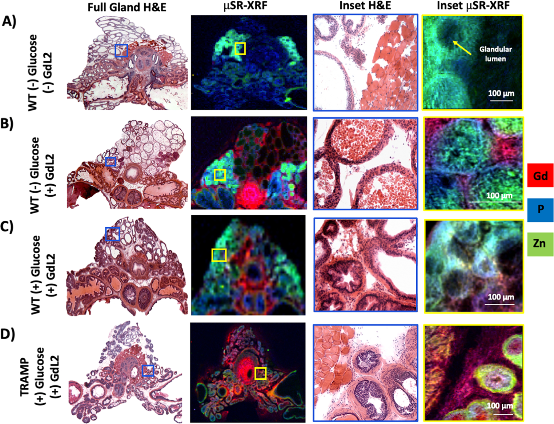
Prostatic zinc content is a known biomarker for discriminating normal healthy tissue from benign prostatic hyperplasia (BPH) and prostate cancer (PCa). Given that zinc content is not readily measured without a tissue biopsy, we have been exploring noninvasive imaging methods to detect these diagnostic differences using a zinc-responsive MRI contrast agent. During imaging studies in mice, we observed that a bolus of glucose stimulates secretion of zinc from the prostate of fasted mice. This discovery allowed the use of a Gd-based zinc sensor to detect differential zinc secretion in regions of healthy versus malignant prostate tissue in a transgenic adenocarcinoma mouse model of PCa. Here, we used a zinc-responsive MRI agent to detect zinc release across the prostate during development of malignancy and confirm the loss of total tissue zinc by synchrotron radiation X-ray fluorescence (μSR-XRF). Quantitative μSR-XRF results show that the lateral lobe of the mouse prostate uniquely accumulates high concentrations of zinc, 1.06 ± 0.08 mM, and that the known loss of zinc content in the prostate is only observed in the lateral lobe during development of PCa. Additionally, we confirm that lesions identified by a loss of zinc secretion indeed represent malignant neoplasia and that the relative zinc concentration in the lesion is reduced to 0.370 ± 0.001 mM. The μSR-XRF data also provided insights into the mechanism of zinc secretion by showing that glucose promotes movement of zinc pools (∼1 mM) from the glandular lumen of the lateral lobe of the mouse prostate into the stromal/smooth muscle surrounding the glands. Co-localization of zinc and gadolinium in the stromal/smooth muscle areas as detected by μSR-XRF confirm that glucose initiates secretion of zinc from intracellular compartments into the extracellular spaces of the gland where it binds to the Gd-based agent and albumin promoting MR image enhancement.
Conflict of interest statement
The authors declare no competing financial interest.
Figures


Similar articles
-
Using micro-synchrotron radiation x-ray fluorescence (µ-SRXRF) for trace metal imaging in the development of MRI contrast agents for prostate cancer imaging.J Trace Elem Med Biol. 2022 Dec;74:127054. doi: 10.1016/j.jtemb.2022.127054. Epub 2022 Aug 3. J Trace Elem Med Biol. 2022. PMID: 35939923 Free PMC article.
-
Zinc-sensitive MRI contrast agent detects differential release of Zn(II) ions from the healthy vs. malignant mouse prostate.Proc Natl Acad Sci U S A. 2016 Sep 13;113(37):E5464-71. doi: 10.1073/pnas.1609450113. Epub 2016 Aug 25. Proc Natl Acad Sci U S A. 2016. PMID: 27562169 Free PMC article.
-
Magnetic Resonance Imaging Detection of Glucose-Stimulated Zinc Secretion in the Enlarged Dog Prostate as a Potential Method for Differentiating Prostate Cancer From Benign Prostatic Hyperplasia.Invest Radiol. 2021 Jul 1;56(7):450-457. doi: 10.1097/RLI.0000000000000760. Invest Radiol. 2021. PMID: 34086013 Free PMC article.
-
Elemental imaging of trace elements in bone samples using micro and nano-X-ray fluorescence spectrometry.Appl Radiat Isot. 2019 Jul;149:200-205. doi: 10.1016/j.apradiso.2019.04.033. Epub 2019 Apr 30. Appl Radiat Isot. 2019. PMID: 31077976 Review.
-
Novel role of zinc in the regulation of prostate citrate metabolism and its implications in prostate cancer.Prostate. 1998 Jun 1;35(4):285-96. doi: 10.1002/(sici)1097-0045(19980601)35:4<285::aid-pros8>3.0.co;2-f. Prostate. 1998. PMID: 9609552 Review.
Cited by
-
Imaging Tissue Physiology In Vivo by Use of Metal Ion-Responsive MRI Contrast Agents.Pharmaceuticals (Basel). 2020 Sep 24;13(10):268. doi: 10.3390/ph13100268. Pharmaceuticals (Basel). 2020. PMID: 32987721 Free PMC article. Review.
-
MRI Methods for Imaging Beta-Cell Function in the Rodent Pancreas.Methods Mol Biol. 2023;2592:101-111. doi: 10.1007/978-1-0716-2807-2_7. Methods Mol Biol. 2023. PMID: 36507988 Free PMC article.
-
From Zn(II) to Cu(II) Detection by MRI Using Metal-Based Probes: Current Progress and Challenges.Pharmaceuticals (Basel). 2020 Nov 30;13(12):436. doi: 10.3390/ph13120436. Pharmaceuticals (Basel). 2020. PMID: 33266014 Free PMC article. Review.
-
Using micro-synchrotron radiation x-ray fluorescence (µ-SRXRF) for trace metal imaging in the development of MRI contrast agents for prostate cancer imaging.J Trace Elem Med Biol. 2022 Dec;74:127054. doi: 10.1016/j.jtemb.2022.127054. Epub 2022 Aug 3. J Trace Elem Med Biol. 2022. PMID: 35939923 Free PMC article.
-
Manganese(II)-Based Responsive Contrast Agent Detects Glucose-Stimulated Zinc Secretion from the Mouse Pancreas and Prostate by MRI.Inorg Chem. 2021 Feb 15;60(4):2168-2177. doi: 10.1021/acs.inorgchem.0c02688. Epub 2021 Jan 28. Inorg Chem. 2021. PMID: 33507742 Free PMC article.
References
-
- Hare DJ; New EJ; de Jonge MD; McColl G Imaging metals in biology: balancing sensitivity, selectivity and spatial resolution. Chem. Soc. Rev 2015, 44 (17), 5941–58. - PubMed
MeSH terms
Substances
Grants and funding
LinkOut - more resources
Full Text Sources
Medical
Research Materials
