. 2019 Jul 1;17(7):e3000326.
doi: 10.1371/journal.pbio.3000326. eCollection 2019 Jul.

Uncoordinated maturation of developing and regenerating postnatal mammalian vestibular hair cells

Affiliations

Uncoordinated maturation of developing and regenerating postnatal mammalian vestibular hair cells

Tian Wang et al. PLoS Biol. .

Abstract

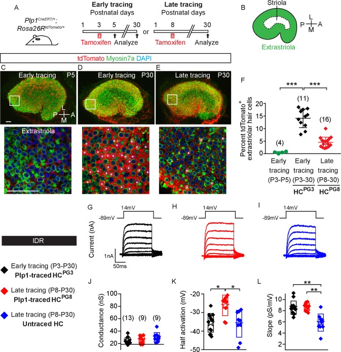
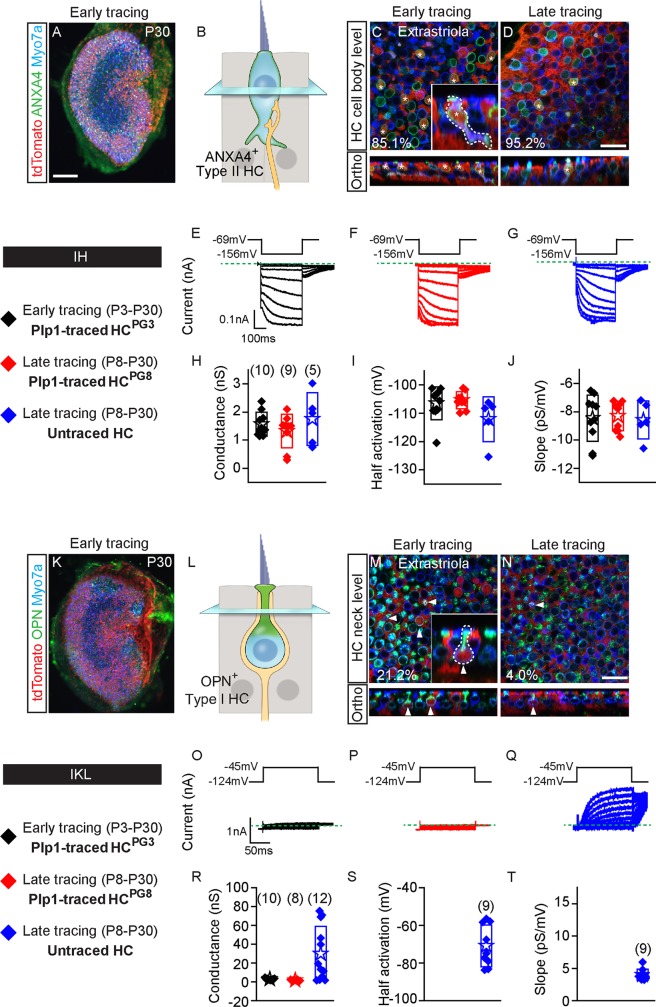
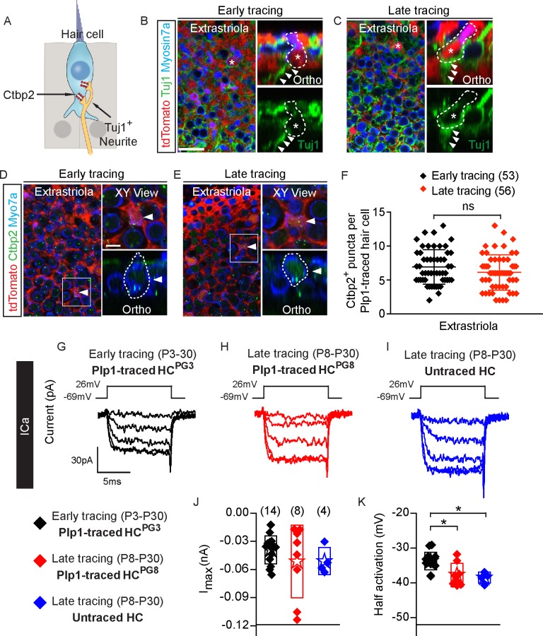
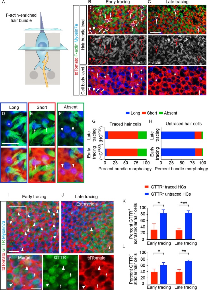
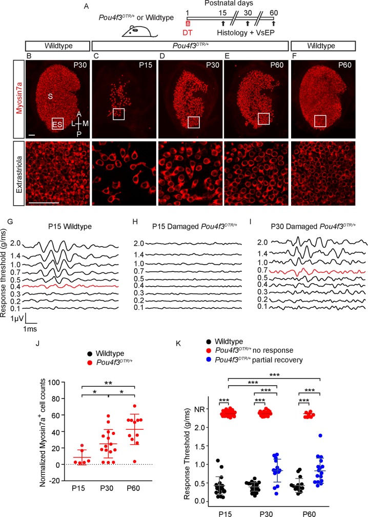
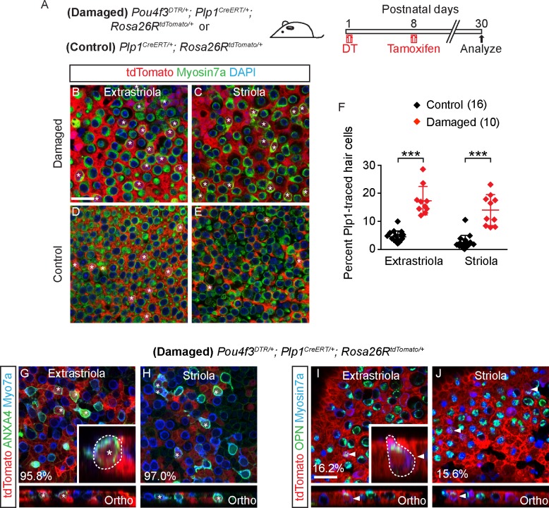
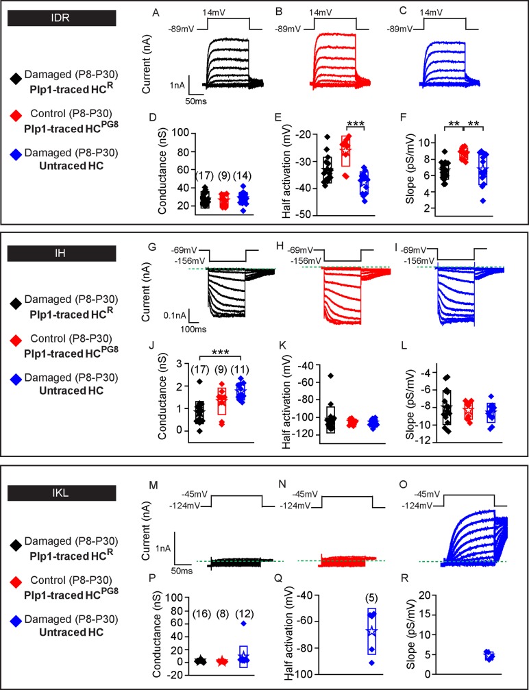
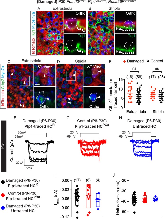
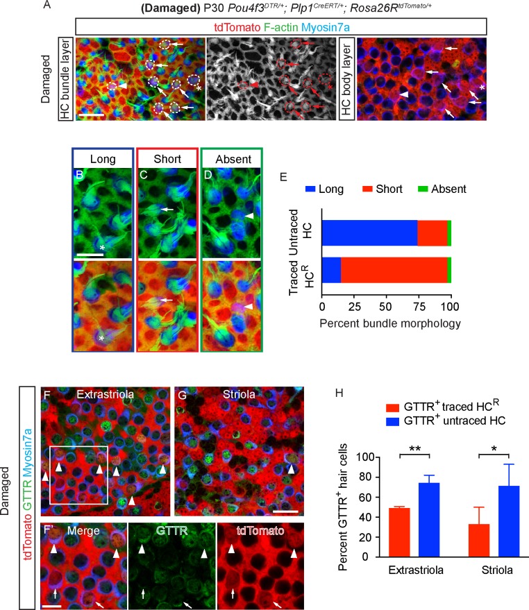
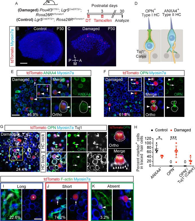
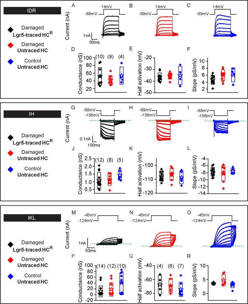
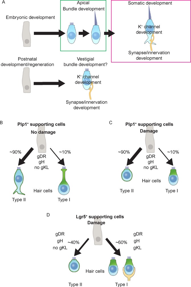
Sensory hair cells are mechanoreceptors required for hearing and balance functions. From embryonic development, hair cells acquire apical stereociliary bundles for mechanosensation, basolateral ion channels that shape receptor potential, and synaptic contacts for conveying information centrally. These key maturation steps are sequential and presumed coupled; however, whether hair cells emerging postnatally mature similarly is unknown. Here, we show that in vivo postnatally generated and regenerated hair cells in the utricle, a vestibular organ detecting linear acceleration, acquired some mature somatic features but hair bundles appeared nonfunctional and short. The utricle consists of two hair cell subtypes with distinct morphological, electrophysiological and synaptic features. In both the undamaged and damaged utricle, fate-mapping and electrophysiology experiments showed that Plp1+ supporting cells took on type II hair cell properties based on molecular markers, basolateral conductances and synaptic properties yet stereociliary bundles were absent, or small and nonfunctional. By contrast, Lgr5+ supporting cells regenerated hair cells with type I and II properties, representing a distinct hair cell precursor subtype. Lastly, direct physiological measurements showed that utricular function abolished by damage was partially regained during regeneration. Together, our data reveal a previously unrecognized aberrant maturation program for hair cells generated and regenerated postnatally and may have broad implications for inner ear regenerative therapies.


Conflict of interest statement

The authors have declared that no competing interests exist.

Figures

Fig 1
Fig 1. Characteristics of postnatally generated hair cells in the mouse utricle.
A) Plp1CreERT/+; Rosa26RtdTomato/+ mice were treated with tamoxifen at P3 (early tracing) and P8 (late tracing) to fate-map supporting cells. Organs were examined at P5 and P30. B) Diagram illustrating the extrastriolar and striolar regions of the utricle. C) Rare tdTomato+/Myosin7a+ hair cells were detected in the extrastriola 2 days after early tracing with tamoxifen given at P3. D) Many traced hair cells (asterisks) were found in the extrastriola at P30. E) After late tracing initiated at P8, few traced hair cells (asterisks) were detected in the extrastriola at P30. Representative high magnification images were selected from the extrastriola region (white boxes). F) Compared to P5, the percentage of tdTomato+/Myosin7a+ cells was significantly higher at P30 after early tracing from P3. It is also significantly higher than that of late tracing from P8 (n = 4–16 mice). G-I) Currents from hair cells were elicited using the delayed rectifier protocol described in the methods. J-L) Conductance-voltage plots were generated and values for maximal conductance, half-activation voltage and slope of the Boltzmann function were extracted (n = 9–13 cells). All examined hair cells from three groups displayed measurable conductances representative of gDR, with some significant differences in half-activation voltage and slope. Data shown as mean ± SD, compared using Student t tests and one-way ANOVA by Kruskal Wallis-Dunn's multiple comparison tests. ***p < 0.001, **p < 0.01, *p < 0.05. Scale bars: 50 μm. The underlying data can be found within S1 Data. P, postnatal day.
Fig 2
Fig 2. Early and late postnatally generated hair cells primarily show type II hair cell characteristics.
A) P30 whole mount utricle (early tracing) labeled for Annexin A4 (ANXA4). B) Cartoon depicting nucleus level of ANXA4+ (green) type II hair cells. C-D) Almost all postnatally generated tdTomato+/Myosin7a+ hair cells from early and late tracing expressed ANXA4 (asterisks). Inset shows orthogonal view of traced goblet-shaped, ANXA4+ type II hair cells with basolateral processes. E-G) Representative tracings of voltage-gated currents in response to the inward rectifier protocol from HCPG3, HCPG8 and untraced hair cells (n = 5–10 cells). H-J) All three groups demonstrate similar inward rectifier electrophysiological (gH) properties: peak conductances, half-activation and slopes. K) P30 utricle from early tracing labeled for OPN. L) Diagram illustrating the nucleus level of OPN+ (green) type I hair cells. Occasional tdTomato+/Myosin7a+ hair cells from early M) and late tracing N) expressed OPN on the apical neck (arrowhead). Inset shows orthogonal view of traced OPN+ type I hair cells with amphora shape, and a long and narrow apical neck. O-Q) Examples of low voltage activated potassium current (IKL) responses during the step displacements for HCPG3, HCPG8 and untraced hair cells (n = 8–12 cells). Green dashed lines define zero current levels. R-T) Similar tail current and reversal potential analysis were performed with resultant data for conductance, half-activation and slope. Data shown as mean ± SD, compared using one-way ANOVA by Kruskal Wallis-Dunn's multiple comparison test. Scale bars: A, K) 100 μm; C-D, M-N) 20 μm. The underlying data can be found within S1 Data. ANXA4, Annexin A4; HCPG, postnatally generated hair cell; OPN, Osteopontin; P, postnatal day.
Fig 3
Fig 3. Synaptic properties of postnatally generated hair cells.
A) Diagram illustrating hair cells with the presynaptic element Ctbp2 (red) and associated Tuj1+ vestibular neurites (beige). B-C) Representative confocal images of tdTomato+/Myosin7a+ hair cells (asterisks and dashed lines) from early and late tracing associated with Tuj1+ neurites (arrowheads in orthogonal views, n = 127 cells from 3 mice for early tracing and 73 cells from 9 mice for late tracing). D-E) Expression of Ctbp2 on the basolateral surfaces of traced hair cells (arrowhead). Shown are high magnification XY and orthogonal views of cells of interests in boxes (n = 53 cells from 3 mice for early tracing and 56 cells from 6 mice for late tracing). F) Quantification of Ctbp2+ puncta in tdTomato+/Myosin7a+ hair cells in the extrastriola. No significant difference was found between early and late tracing. G-I) Representative calcium currents from Plp1-traced HCPG3, HCPG8 and untraced hair cells. Calcium currents were isolated from voltage-clamped hair cells by replacing intracellular potassium with Cs and tetra-ethyl ammonium. Currents in response to the calcium current protocol were monitored and analyzed. J-K) Current-voltage plots were generated from peak current responses and maximal current and half activating voltage were extracted from these plots (n = 4–14 cells). Data shown as mean ± SD, compared using Student t tests and one-way ANOVA by Kruskal Wallis-Dunn's multiple comparison test. *p < 0.05. Scale bars: B-E) 20 μm, XY views in D-E) 5 μm. The underlying data can be found within S1 Data. HCPG, postnatally generated hair cell.
Fig 4
Fig 4. Bundle features of early and late postnatally generated hair cells.
A) Diagram illustrating hair cell stereociliary bundles. B-C) tdTomato+/Myosin7a+ hair cells (dashed circles) added from early and late tracing with long (asterisks), short (arrows) and absent bundles (arrowheads) in both the hair bundle and cell body level. D-F) High magnification images of representative cells with long (D, asterisks), short (E, arrows) and absent bundles (F, arrowheads). G-H) Proportion of bundle morphology in traced and untraced hair cells. The majority of traced hair cells had short bundles from early (n = 127 HCs from 4 mice) and late tracing (n = 96 from 8 mice), whereas majority of untraced hair cells had long bundles (n = 849 HCs from 4 mice at P3, 794 HCs from 8 mice at P8). I-J) Representative confocal images of GTTR+/tdTomato+/Myosin7a+ (arrowhead) and GTTR-/tdTomato+/Myosin7a+ (arrows) hair cells from early and late tracing. I’) High magnification images of panel I. K-L) Significantly more untraced hair cells than traced hair cells were GTTR-labeled in both early (n = 117 and 48 traced HCs, 649 and 700 untraced HCs from 3 mice in the extrastriola and striola) and late tracing experiments (n = 30 and 16 traced HCs, 951 and 740 untraced HCs from 4 mice in the extrastriola and striola). Data shown as mean ± SD, compared using Student t tests. ***p < 0.001, **p < 0.01, *p < 0.05. Scale bars: B-C, I-J) 20 μm; D-F, I’) 10 μm. The underlying data can be found within S1 Data. P, postnatal day.
Fig 5
Fig 5. Hair cell number and vestibular function recovers after hair cell ablation.
A) Pou4f3DTR/+ mice were treated with DT at P1 and examined at P15 and P30 and P60. B-F) After DT treatment, both the hair cell number and area of sensory epithelium decreased at P15, followed by a partial recovery at P30 and P60. G-I) Representative VsEP waveforms of normal responses of a P15 wild-type animal (0.4 g/ms threshold shown in red) (G), absent responses in a damaged P15 Pou4f3DTR/+ mouse (H), and an elevated response threshold in a damaged P30 Pou4f3DTR/+ mouse (0.7 g/ms threshold shown) (I). J) Normalized Myosin7a+ hair cell counts at P30 are significantly higher than P15 (n = 6 at P15 and 15 at P30). Damaged tissues normalized to age-matched controls. K) Many animals displayed partial VsEP thresholds recovery (blue dots) at P30 and P60 although some still showed no responses (red dots). Compared to P15, the average thresholds at P30 and P60 are significantly lower (n = 48 at P15, 39 at P30 and 27 at P60), but still significantly higher than age-matched controls (n = 20 at P15, 22 at P30, and 15 at P60). Data shown as mean ± SD, compared using Student t tests and one-way ANOVA by Tukey’s multiple comparison test. ***p < 0.001, **p < 0.01, *p < 0.05. Scale bars: 50 μm in B-F. The underlying data can be found within S1 Data. DT, diphtheria toxin; P, postnatal day; VsEP, vestibular-evoked potential.
Fig 6
Fig 6. Plp1+ supporting cells regenerate hair cells after hair cell loss.
A) Pou4f3DTR/+; Plp1CreERT/+; Rosa26RtdTomato/+ mice were treated with DT at P1, followed by tamoxifen at P8 to fate-map Plp1+ supporting cells. Organs were examined at P30. B-E) Damaged P30 utricles had more tdTomato+/Myosin7a+ hair cells (asterisks) than controls. F) Quantification shows that the percentage of tdTomato+/Myosin7a+ hair cells in damaged utricles (n = 10) was significantly higher than in undamaged controls (n = 16). G-H) Almost all traced, regenerated hair cells (asterisks) expressed the type II hair cell marker ANXA4 in both the extrastriola and striola. Inset shows orthogonal view of a traced, regenerated ANXA4+ hair cell with a short, round cell body and no basolateral processes. I-J) Occasionally traced, regenerated hair cells (arrowhead) expressed the type I hair cell marker OPN in both the extrastriola and striola. Inset shows orthogonal view of traced, regenerated OPN+ hair cells appearing pear-shaped, with a short neck. Data shown as mean ± SD and compared using Student t tests. ***p < 0.001. Scale bars: 20 μm. The underlying data can be found within S1 Data. ANXA4, Annexin A4; OPN, Osteopontin.
Fig 7
Fig 7. Plp1-traced, regenerated hair cells display electrophysiological features of type II hair cells.
Three groups of hair cells were examined: 1) traced (black, HCR) and 2) untraced hair cells (blue) from P30 Pou4f3DTR/+; Plp1CreERT/+; Rosa26RtdTomato/+ mice treated with DT at P1 and tamoxifen at P8; and 3) traced hair cells (red, HCPG8) from Plp1CreERT/+; Rosa26RtdTomato/+ mice treated and tamoxifen at P8. A-C) Representative tracings of IDR from hair cells from each group (HCR, HCPG8 and untraced hair cells, n = 9–17 cells). D-F) All three groups demonstrate similar peak conductances but significant differences between half-activation and slopes. G-I) Tracings of IH measurements from HCR, HCPG8 and untraced hair cells (n = 9–17 cells). J-L) All three groups demonstrate similar peak conductances, half-activation, and slopes. M-O) IKL responses from untraced hair cells from the damaged utricles, but not HCR or HCPG8 (n = 8–16 cells). P-R) Similar tail current and reversal potential analysis were performed and the resultant data for conductance, half-activation and slope for untraced hair cells are presented. Data shown as mean ± SD and compared using one-way ANOVA by Kruskal Wallis-Dunn's multiple comparison tests. Green dashed lines define zero current levels. ***p < 0.001, **p < 0.01. The underlying data can be found within S1 Data. HCPG, postnatally generated hair cell; P, postnatal day.
Fig 8
Fig 8. Synaptic properties of Plp1-traced, regenerated hair cells.
A-B) Representative images of regenerated tdTomato+/Myosin7a+ hair cells (asterisks) with associated with Tuj1+ neural elements (arrowheads) in the extrastriola and striola (n = 106 cells from 5 mice). C-D) All Plp1-traced hair cells examined expressed Ctbp2 on the basolateral surfaces (arrowheads, n = 35 cells from 3 mice). E) Quantification of Ctbp2+ puncta in traced hair cells in the extrastriola and striola. No significant difference was found between hair cells from control and damaged utricles (n = 56 extrastriolar and 25 striolar hair cells from 6 control mice utricles, n = 18 extrastriolar and 17 striolar hair cells from 3 damaged mice utricles). F-H) Representative calcium currents from traced hair cells (HCR from damaged utricles and HCPG8 from undamaged utricles) and untraced hair cells. Calcium currents were isolated using methods described in Fig 3. I-J) Peak current responses and maximal current and half activating voltage were not statistically different among groups (n = 4–17 cells). Data shown as mean ± SD, compared using Student t tests and one-way ANOVA by Kruskal Wallis-Dunn's multiple comparison tests. Scale bars: A-D) 20 μm, XY view) 5 μm. The underlying data can be found within S1 Data. HCPG, postnatally generated hair cell.
Fig 9
Fig 9. Regenerated hair cells display immature stereociliary bundles.
A) Regenerated traced hair cells (dashed circles) with long (asterisks), short (arrows) and absent bundles (arrowheads). Shown are images taken at the focal planes of hair bundle and hair cell body. B-D) Representative high magnification images of hair cells with long (B) (asterisk), short (C) (arrow) and absent bundles (D) (arrowhead). E) Proportion of traced and untraced hair cells displaying the above bundle morphology. Most regenerated hair cells had short bundles (124 cells from 6 damaged mice), whereas most untraced hair cells had long bundles (617 cells from 6 damaged mice). F-G) Representative confocal images of GTTR+/tdTomato+/Myosin7a+ (arrowhead) and GTTR-/tdTomato+/Myosin7a+ hair cells (arrows) in damaged utricles. F’) High magnification images of panel F. H) Compared to traced hair cells, significantly more untraced hair cells were GTTR-labeled in the damaged utricles (n = 25 and 11 traced HCs, 172 and 148 untraced HCs from 4 mice in the extrastriola and striola). Data shown as mean ± SD, compared using Student t tests. **p < 0.01, *p < 0.05. Scale bars: A, F-G) 20 μm; B-D, F’) 10 μm. The underlying data can be found within S1 Data.
Fig 10
Fig 10. Lgr5+ supporting cells regenerate type I and type II hair cells.
A) Pou4f3DTR/+; Lgr5CreERT2/+; Rosa26RtdTomato/+ and Lgr5CreERT2/+; Rosa26RtdTomato/+ mice were treated with DT at P1, followed by tamoxifen at P3 to fate-map Lgr5+ supporting cells. Organs were examined at P30. B-C) Whole mount preparation of P30 control and damaged utricles. Traced cells primarily occupied the presumed striolar region of the damaged organ. D) Diagram illustrating type I and II hair cells, the former of which are endowed with Tuj1+ calyx. E) Representative images of ANXA4+ (arrowhead) and ANXA4- (arrow) traced hair cells in the P30 damaged utricle. 46.9% of Lgr5-traced hair cells expressed ANXA4 (n = 36 cells from 3 mice). F) Representative images of OPN+ (arrowhead) and OPN- (arrow) traced hair cells in the P30 damaged utricle. 61.8% of Lgr5-traced hair cells were OPN+ (n = 71 cells from 7 mice). G) In the P30 damaged utricle, Lgr5-traced hair cells expressed OPN (arrowhead) and were surrounded by Tuj1+ calyx (arrowhead, 24.4% of Lgr5-traced hair cells, n = 40 cells from 4 mice), or were OPN- and innervated by Tuj1+ boutons (arrow). Representative orthogonal view shows round, pear-shaped, regenerated hair cells with OPN+, short neck and Tuj1+ calyx (arrowheads). H) Compared to undamaged controls, the damages utricles had significantly fewer ANXA4+ traced HCR and more OPN+ and OPN+/Tuj1+ (Calyx) traced HCR. No OPN+ and OPN+/Tuj1+ (Calyx) traced hair cells were seen in the undamaged utricle. Data shown as mean ± SD, compared using Student t tests. (I-K) Representative high magnification images of traced hair cells with long (I) (asterisk), short (J) (arrow) and absent bundles (K) (arrowhead). ***p < 0.001, *p < 0.05. Scale bars: B-C) 100 μm; E-G) 20 μm. (I-K) 10 μm. The underlying data can be found within S1 Data. ANXA4, Annexin A4; OPN, Osteopontin.
Fig 11
Fig 11. Lgr5-traced, regenerated hair cells display electrophysiological features of type II and type I hair cells.
Three groups of hair cells were examined: 1) traced (black, HCR) and 2) untraced (red) hair cells from P30 Pou4f3DTR/+; Lgr5CreERT2/+; Rosa26RtdTomato/+ mice treated with DT at P1 and tamoxifen at P3; and untraced (blue) hair cells from Lgr5CreERT2/+; Rosa26RtdTomato/+ mice treated with tamoxifen at P3. A-C) Representative tracings of IDR from hair cells from each group (HCR, untraced hair cells from damage and controls, n = 4–10 cells). D-F) All three groups demonstrate similar peak conductances, half-activation and slopes. G-I) Tracings of IH measurements from HCR, and untraced hair cells from damaged and undamaged organs (n = 5–12 cells). J-L) All three groups demonstrate similar electrophysiological properties: peak conductances, half-activation and slopes. M-O) IKL responses from HCR, and untraced hair cells from damaged and undamaged organs (n = 10–14 cells). P-R) Conductance, half-activation and slopes were no different among the three groups. Data shown as mean ± SD, compared using one-way ANOVA by Kruskal Wallis-Dunn's multiple comparison tests. Green dashed lines define zero current levels. The underlying data can be found within S1 Data.
Fig 12
Fig 12. Maturation pathways of developing and regenerating hair cells.
A) Vestibular hair cells specified early during embryonic period acquire stereocilia, differentiate into subtypes of hair cells, and subsequent innervation. Postnatally generated/regenerated hair cells develop somatic features including differentiation into hair cell subtypes and innervation but were delayed in apical bundle development. B) In the undamaged utricle, majority of Plp1+ supporting cells generate type II hair cells, with type I hair cells are only occasionally added. C) In the damaged utricle, majority of Plp1+ supporting cells regenerate type II hair cells, type I hair cells are occasionally regenerated. D) By contrast, in the damaged utricle, Lgr5+ supporting cells regenerate a similar ratio of type II and type I hair cells. A subset of type I HCR was innervated with Tuj1+ calyx (24.4%).

Similar articles

Cited by

References

    1. Bermingham-McDonogh O, Reh TA Regulated reprogramming in the regeneration of sensory receptor cells. Neuron, (2011); 71: 389–405. 10.1016/j.neuron.2011.07.015 - DOI - PMC - PubMed
    1. Burns JC, Cox BC, Thiede BR, Zuo J, Corwin JT In vivo proliferative regeneration of balance hair cells in newborn mice. J Neurosci, (2012); 32: 6570–6577. 10.1523/JNEUROSCI.6274-11.2012 - DOI - PMC - PubMed
    1. Forge A, Li L, Corwin JT, Nevill G Ultrastructural evidence for hair cell regeneration in the mammalian inner ear. Science, (1993); 259: 1616–1619. - PubMed
    1. Forge A, Li L, Nevill G Hair cell recovery in the vestibular sensory epithelia of mature guinea pigs. J Comp Neurol, (1998); 397: 69–88. - PubMed
    1. Golub JS, Tong L, Ngyuen TB, Hume CR, Palmiter RD, Rubel EW, et al. Hair cell replacement in adult mouse utricles after targeted ablation of hair cells with diphtheria toxin. J Neurosci, (2012); 32: 15093–15105. 10.1523/JNEUROSCI.1709-12.2012 - DOI - PMC - PubMed

Publication types

MeSH terms

LinkOut - more resources