. 2019 Nov;106(5):1139-1151.
doi: 10.1002/JLB.3A0119-025RRR. Epub 2019 Jul 1.

Ablation of RhoA impairs Th17 cell differentiation and alleviates house dust mite-triggered allergic airway inflammation

Affiliations

Ablation of RhoA impairs Th17 cell differentiation and alleviates house dust mite-triggered allergic airway inflammation

Jun-Qi Yang et al. J Leukoc Biol. 2019 Nov.

Abstract

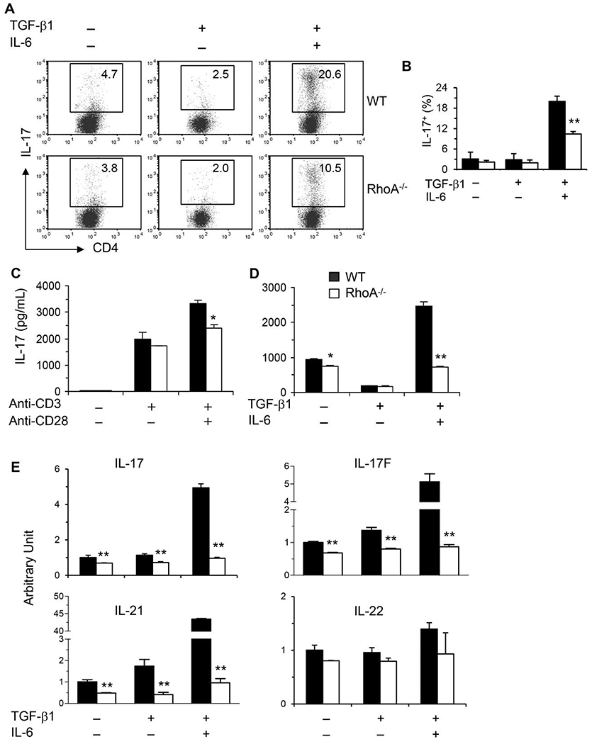
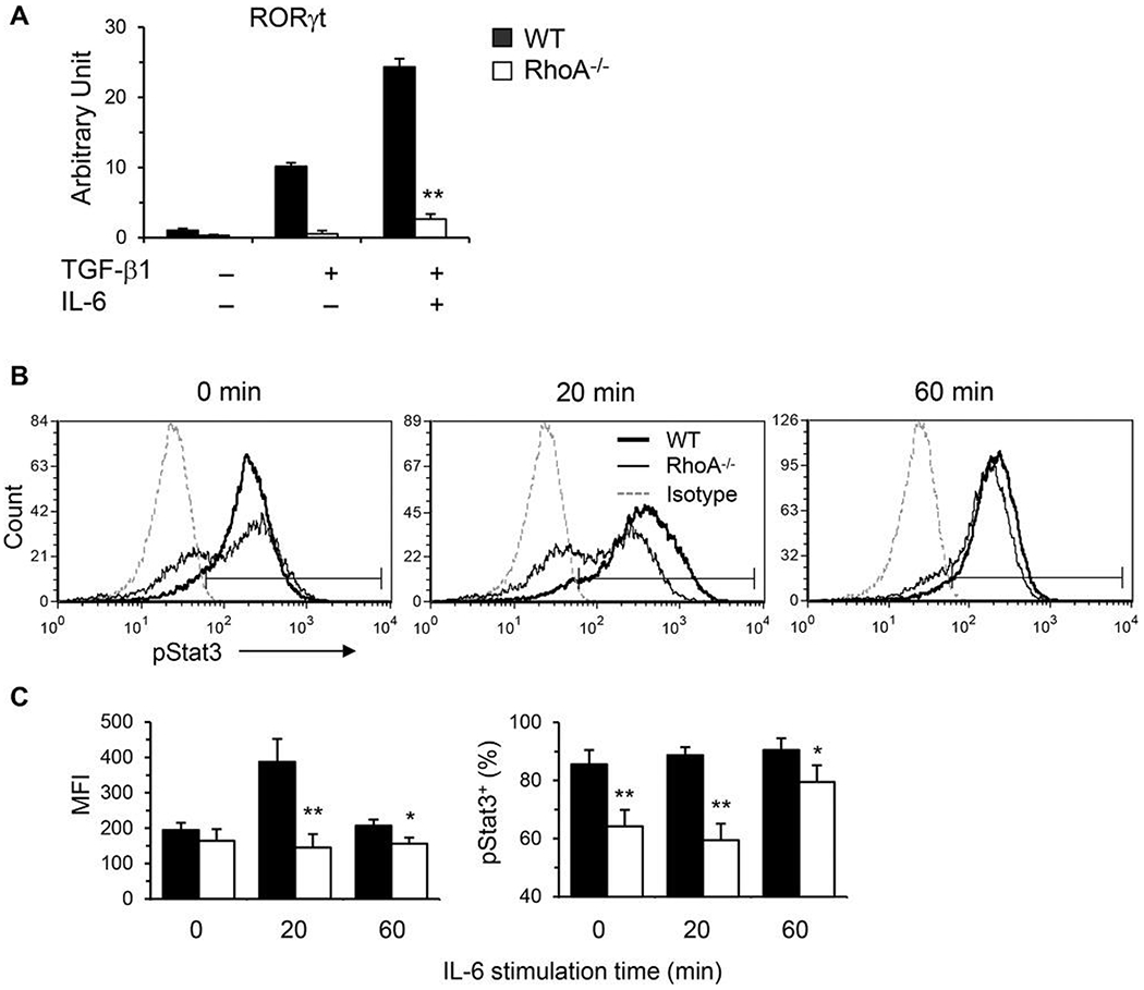
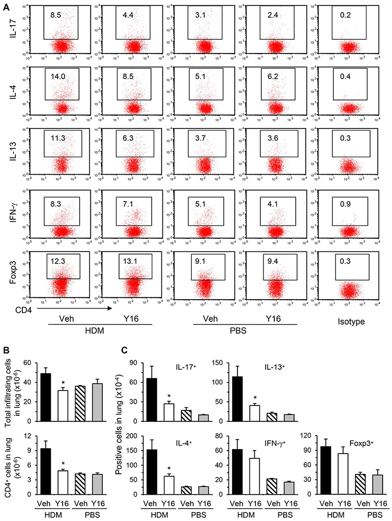
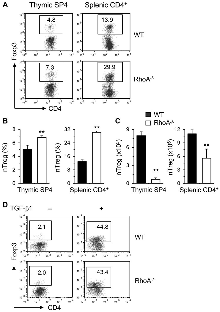
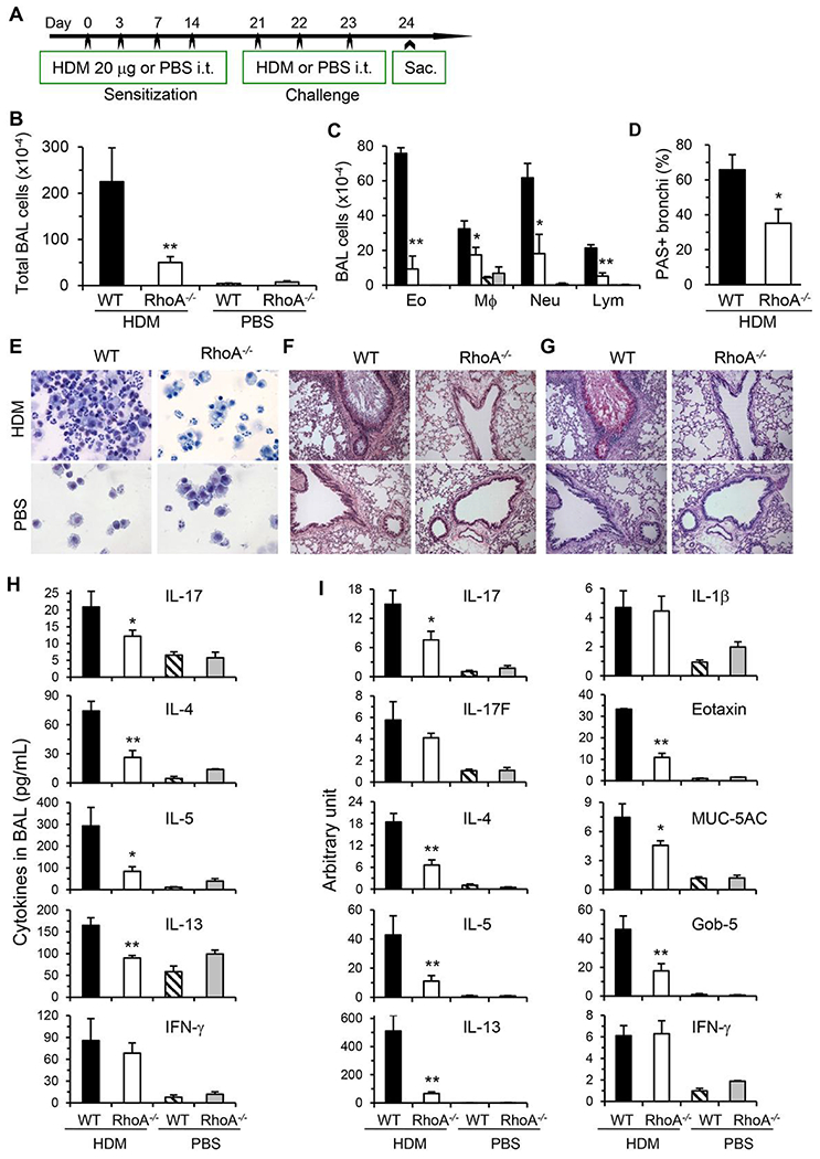
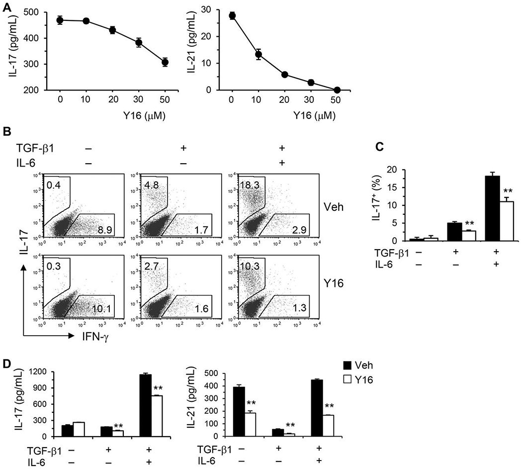
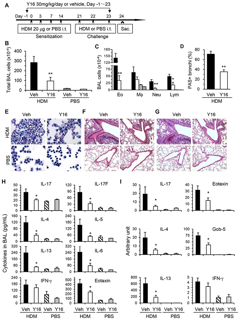
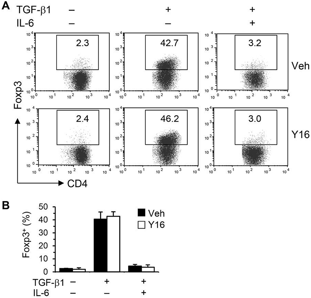
Asthma is a heterogeneous chronic airway inflammation in which Th2 and Th17 cells are key players in its pathogenesis. We have reported that RhoA of Rho GTPases orchestrated glycolysis for Th2 cell differentiation and allergic airway inflammation by the use of a conditional RhoA-deficient mouse line. However, the role of RhoA in Th17 cells remains to be elucidated. In this study, we investigated the effects of RhoA deficiency on Th17 cells in the context of ex vivo cell culture systems and an in vivo house dust mites (HDM)-induced allergic airway inflammation. We found that RhoA deficiency inhibited Th17 differentiation and effector cytokine secretion, which was associated with the downregulations of Stat3 and Rorγt, key Th17 transcription factors. Furthermore, loss of RhoA markedly suppressed Th17 and neutrophil-involved airway inflammation induced by HDM in mice. The infiltrating inflammatory cells in the lungs and bronchoalveolar lavage (BAL) fluids were dramatically reduced in conditional RhoA-deficient mice. Th17 as well as Th2 effector cytokines were suppressed in the airways at both protein and mRNA levels. Interestingly, Y16, a specific RhoA inhibitor, was able to recapitulate the most phenotypes of RhoA genetic deletion in Th17 differentiation and allergic airway inflammation. Our data demonstrate that RhoA is a key regulator of Th17 cell differentiation and function. RhoA might serve as a potential novel therapeutic target for asthma and other inflammatory disorders.

Keywords: RhoA; Th17; Y16; allergic airway inflammation; house dust mite.


Conflict of interest statement

CONFLICTS OF INTEREST

The authors declare no conflicts of interest.

Figures

FIGURE 1
FIGURE 1. RhoA deficiency dampens Th17 differentiation.
(A and B) Purified CD4+ T cells were differentiated under Th17-polarizing conditions (TGF-β1 + IL-6) for 4 days and restimulated with PMA plus ionomycin for 5 h with BD GolgiPlug in the last 2 h. Cells were collected for IL-17 intracellular staining. Percentages of IL-17+ cells are shown in representative dot plots (A) and summarized in a bar graph (B). (C and D) Supernatants were collected from CD4+ T cells stimulated with or without anti-CD3/CD28 mAbs (C) or differentiated under Th17-polarizing conditions and restimulated with PMA/ionomycin (D)for ELISA assays of IL-17. (E) Total RNA was extracted from the cultured cells for real-time PCR analysis. The mRNA levels of Th17 effector cytokines as indicated are shown (arbitrary unit). The data are normalized to an 18S reference. Results (B-E) are expressed as mean + SD of triplicates, representative of 3 independent experiments. *P < 0.05, **P < 0.01 compared to WT cells
FIGURE 2
FIGURE 2. RhoA deficiency downregulates Th17 transcription factors.
(A) Total RNA was extracted from splenic CD4+ T cells differentiated under Th17 conditions and restimulated with PMA/ionomycin for real-time PCR analysis. The mRNA levels of RORγt are shown. The data are normalized to an 18S reference. (B and C) Naïve splenic CD4+ T cells were stimulated with plate-bound anti-CD3 in the presence of free anti-CD28 for 3 days. Cells were washed and restimulated with recombinant mouse IL-6 (25 ng/mL) for the indicated time. Cells were harvested and processed for intracellular FACS staining for pStat3 (pY705) following BD Bioscience’s protocols. Representative histograms (B), the mean fluorescence intensity (MFI) and percentages of pStat3+ cells (C) are shown. Naïve CD4+ T cells were pooled from 5 to 6 mice. Results (means + SD of triplicates) are representative of 2 independent experiments. *p < 0.05, **P < 0.01 versus WT cells
FIGURE 3
FIGURE 3. Effect of RhoA deficiency on Treg cells.
(A-C) Total thymocytes and splenocytes were prepared from conditional RhoA-deficient mice and their WT littermates. Cells were processed for Treg staining. Percentages of natural Treg (nTreg, Foxp3+) in thymic single CD4+(SP4) and splenic CD4+ cells are shown in representative dot plots (A) and summarized in bar graphs (B). The absolute numbers of nTreg are also shown (C) (mean + SD, n = 5). (D) Naive CD4+ T cells were differentiated under Treg polarizing condition with TGF-β1 for 4 days and restimulated with PMA plus ionomycin. Percentages of Foxp3+ cells are shown in representative dot plots. Data are representative of 2 independent experiments with similar results. **P < 0.01 compared to WT mice
FIGURE 4
FIGURE 4. RhoA deficiency inhibits HDM-induced allergic airway inflammation.
(A) RhoA−/− mice and their WT littermates were inoculated and challenged intratracheally (i.t.) with HDM to induce allergic airway inflammation. Control groups were inoculated i.t. with PBS alone. Mice were sacrificed 24 h after the last challenge. (B and C) Quantification of total BAL cells (B), eosinophils (Eo), macrophages (Mø), neutrophils (Neu), and lymphocytes (Lym) (C) in BAL fluids. (D-G) Representative Kwik-Diff staining for BAL cytospins (E), H&E (F), and PAS staining (G) of same areas in lung tissue sections. Percentages of PAS-stained bronchi are summarized (D). (H) Cytokine and chemokine levels in BAL fluids determined by ELISA. (I) mRNA levels of cytokines and chemokines in lung tissues determined by real-time PCR. Data are normalized to an 18S reference and expressed as arbitrary units. Results (means + SE, n = 6-8) are representative of 2 independent experiments. *P < 0.05, **P < 0.01 versus HDM-challenged WT groups
FIGURE 5
FIGURE 5. RhoA specific inhibitor Y16 blocks Th17 differentiation.
(A) Purified naïve CD4+ T cells (1 × 10⁶/mL) pooled from 8 WT mice were stimulated with plate-bound anti-CD3 plus free anti-CD28 for 2 days without or with Y16 (0~50 μM). IL-17 and IL-21 in the culture supernatants were determined by ELISA. (B-D) Naïve CD4+ T cells were differentiated under Th17 conditions for 4 days and restimulated with PMA plus ionomycin for 5 h, in the presence of vehicle (Veh) or Y16 (30 μM) throughout the culture. Cells were collected for IL-17/IFN-γ intracellular staining. Percentages of IL-17+ or IFN-γ+ CD4+ T cells are shown in representative dot plots (B) and summarized in a bar graph (C). IL-17 and IL-21 in the culture supernatants were determined by ELISA (D). Data are representative of 2 independent experiments. **P < 0.01 compared to vehicle-treated groups
FIGURE 6
FIGURE 6. Y16 has no effect on Treg induction in vitro.
Naïve CD4+ T cells were differentiated under Treg/Th17 conditions for 4 days and restimulated with PMA plus ionomycin for 5 h, in the presence of vehicle (Veh) or Y16 (30 μM) throughout the culture. Cells were collected for Treg staining. Percentages of Foxp3+ CD4+ T cells are shown in representative dot plots (A) and summarized in a bargraph (B). Data are representative of 2 independent experiments
FIGURE 7
FIGURE 7. Y16 alleviates HDM-induced allergic airway inflammation.
WT mice were injected intraperitoneally (i.p.) daily with Y16 (30 mg/kg) or vehicle (Veh), starting 1 day before HDM inoculation until 1 day before sacrifice (A). Total cells (B) and differential cell counts (C) in BAL fluids, representative Kwik-Diff staining for BAL cytospins (E), H&E (F), and PAS (G) staining of same areas in lung tissue sections are shown. Percentages of PAS stained bronchi are summarized (D). Cytokine and chemokine levels in BAL fluids (H), and their mRNA levels in lung tissues determined by real-time PCR (I) are shown. Results (means + SE, n = 4-8) are representative of 2 independent experiments. *P < 0.05, **P < 0.01 versus HDM-challenged vehicle-treated groups
FIGURE 8
FIGURE 8. Y16 treatment downregulates pulmonary Th17 and Th2 cells from HDM-inoculated mice.
Allergic airway inflammation was induced by HDM and treated with Y16 as described in Fig. 7A. Mice were sacrificed 24 h after the last HDM challenge and the pulmonary infiltrating cells were isolated. The cells were restimulated in vitro with PMA plus ionomycin for 4 h with BD GolgiPlug in the last 3 h for FACS stainings. Percentages of cytokine or Foxp3 positive cells gated on CD4+ T cells are shown in representative dot plots (A). The isotype controls from pooled cells are also shown (A, Right). The numbers of total pulmonary infiltrating cells and CD4+ T cells (B), cytokine or Foxp3 positive cells gated on CD4+ cells (C) are summarized in bar graphs (means + SE, n = 3-6). *P < 0.05 versus HDM-challenged vehicle-treated groups

Similar articles

Cited by

References

    1. Mulloy JC, Cancelas JA, Filippi MD, Kalfa TA, Guo F, Zheng Y. Rho GTPases in hematopoiesis and hemopathies. Blood. 2010;115: 936–947. - PMC - PubMed
    1. Zhou X, Florian MC, Arumugam P, et al. RhoA GTPase controls cytokinesis and programmed necrosis of hematopoietic progenitors. J Exp Med. 2013;210:2371–2385. - PMC - PubMed
    1. Loirand G Rho kinases in health and disease: from basic science to translational research. Pharmacol Rev. 2015;67:1074–1095. - PubMed
    1. Li H, Peyrollier K, Kilic G, Brakebusch C. Rho GTPases and cancer. Biofactors. 2014;40:226–235. - PubMed
    1. Henning SW, Galandrini R, Hall A, Cantrell DA. The GTPase Rho has a critical regulatory role in thymus development. Embo J. 1997;16: 2397–2407. - PMC - PubMed

Publication types

MeSH terms

Substances