Review
. 2019 Oct 4;431(21):4290-4320.
doi: 10.1016/j.jmb.2019.06.029. Epub 2019 Jun 29.

Distinct Genome Replication and Transcription Strategies within the Growing Filovirus Family

Affiliations
Review

Distinct Genome Replication and Transcription Strategies within the Growing Filovirus Family

Adam J Hume et al. J Mol Biol. .

Abstract

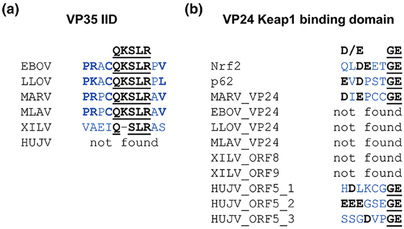
Research on filoviruses has historically focused on the highly pathogenic ebola- and marburgviruses. Indeed, until recently, these were the only two genera in the filovirus family. Recent advances in sequencing technologies have facilitated the discovery of not only a new ebolavirus, but also three new filovirus genera and a sixth proposed genus. While two of these new genera are similar to the ebola- and marburgviruses, the other two, discovered in saltwater fishes, are considerably more diverse. Nonetheless, these viruses retain a number of key features of the other filoviruses. Here, we review the key characteristics of filovirus replication and transcription, highlighting similarities and differences between the viruses. In particular, we focus on key regulatory elements in the genomes, replication and transcription strategies, and the conservation of protein domains and functions among the viruses. In addition, using computational analyses, we were able to identify potential homology and functions for some of the genes of the novel filoviruses with previously unknown functions. Although none of the newly discovered filoviruses have yet been isolated, initial studies of some of these viruses using minigenome systems have yielded insights into their mechanisms of replication and transcription. In general, the Cuevavirus and proposed Dianlovirus genera appear to follow the transcription and replication strategies employed by the ebola- and marburgviruses, respectively. While our knowledge of the fish filoviruses is currently limited to sequence analysis, the lack of certain conserved motifs and even entire genes necessitates that they have evolved distinct mechanisms of replication and transcription.

Keywords: Ebola virus; Marburg virus; filoviruses; genome replication and transcription; nonsegmented negative sense RNA viruses.


Conflict of interest statement

Declarations of interest: none

Figures

Fig. 1.
Fig. 1.. Comparison of filovirus genomes.
(a) A phylogenic tree of the indicated filoviruses was performed by aligning the L protein sequences using the Geneious Prime software. Labeled arcs indicate the officially assigned filovirus genera [6], with the exception of the “Dianloviruses” genus which has been proposed but not yet accepted. BOMV was added to the Ebolavirus genus, although it has not been assigned officially to the genus, (b) Schematics of filovirus genomes. Each filovirus genus is represented by the prototypical virus. Schematics are depicted to scale. Leader and trailer sequences are indicated by black bars, with missing sequence information indicated by squiggly lines. Genes are depicted as colored bars with lighter portions indicating open reading frames (ORFs). Homologous genes are colored similarly. Since the LLOV VP24 and L genes appear to form a bicistronic transcript, the untranslated region separating the VP24 and L ORFs is colored by stripes. For XILV and HUJV, genes with no homology to genes of other filoviruses and no known function are indicated in brown with lighter portions indicating ORFs. Sites of gene overlaps are indicated by black arrows. Intergenic regions are in white. Sites of cotranscriptional mRNA editing are indicated by asterisks. BDBV, Bundibugyo virus; BOMV, Bombali virus; EBOV, Ebola virus; GP, glycoprotein; HUJV, Huángjiāo virus; L, large protein; LLOV, Lloviu virus; MARV, Marburg virus; MLAV, Měnglà virus; NP, nucleoprotein; ORE, open reading frame; RAW, Ravn virus; RESTV, Reston virus; sGP, soluble glycoprotein; ssGP, small soluble glycoprotein; SUDV, Sudan virus; TAFV, Taï Forest virus; XILV, Xīlăng virus.
Fig. 2.
Fig. 2.. Scheme of filovirus particle.
The cell membrane-derived envelope (outer gray oval) is studded with viral glycoprotein trimers (yellow lollipops). The assembly of the matrix protein VP40 (blue) into long, filamentous particles imparts the filovirus particles with their namesake virion shape. The viral nucleocapsid consists of the negative sense genome (black line) enwrapped in the nucleoprotein (NP, pink spheres) along with the polymerase cofactor VP35 (green spheres), the transcription factor VP30 (dark gray spheres), the nucleocapsid protein VP24 (red spheres), and the catalytic polymerase subunit L (light gray ring with appendices). Based on the structure of the vesicular stomatitis virus L protein [132], the filoviral L is hypothetically shown as a ring with an appendage formed by three globular domains.
Fig. 3.
Fig. 3.. Hypothetical model of Ebola virus genome replication and transcription.
(1) EBOV nucleocapsids are released into the cytoplasm of the infected cells after fusion of the viral and the endosomal membranes. The incoming nucleocapsids consist of the NP-RNA complex (NP, pink spheres), VP35 (green spheres), L (light gray ring with appendices), VP24 (red spheres), and contain mostly phosphorylated EBOV VP30 (dark gray spheres). Based on the structure of the vesicular stomatitis virus L protein [132], EBOV L is hypothetically shown as a ring with an appendage formed by three globular domains. (2) The nucleocapsids are released into the cytoplasm of the infected cells where primary transcription occurs. The viral RNA-dependent RNA polymerase formed by the catalytic polymerase subunit L and the cofactor VP35 initiates mRNA synthesis. VP35 binds to NP and redirects L to the NP-RNA complex. VP30 acts as a transcription activator and must be dephosphorylated to be functional. Dephosphorylation of VP30 is carried out by cellular phosphatases (dark blue sphere) interacting with the nucleocapsid. Since VP24 condensates the nucleocapsids, we hypothesize that transcription-competent nucleocapids contain no or only small amounts of VP24. (3) L caps the 5’ ends of the mRNAs and methylates the caps. Polyadenylation of the nascent mRNAs occurs through polymerase stuttering. The GP mRNA is cotranscriptionally edited by polymrase stuttering as indicated by the “+A” in the GP transcript. Dissociation of the viral polymerase at the gene borders results in a transcript gradient. (4) The viral mRNAs are translated by the cellular translation machinery. (5) This enables secondary transcription that requires newly synthesized viral proteins. (6) Phosphorylation of VP30 diminishes its function in viral transcription and helps to initiate viral genome replication. VP40 (blue) and VP24 inhibit viral RNA synthesis. VP35 serves as a chaperone to prevent NP from aggregation. Genome replication involves the synthesis of a positive sense antigenome (blue line) that serves as a replication intermediate. (7) VP24 is involved in nucleocapsid condensation and shifts the viral replication cycle towards assembly and budding. Binding of phosphorylated VP30 to NP ensures packaging of VP30 into virions. Fully assembled nucleocapsids are transported by an actin-dependent transport mechanism to the budding sites. Note that there are signficant differences to Marburg virus transcription as explained in the text.
Fig. 4.
Fig. 4.. Tools to study filovirus replication and transcription.
(a) Filovirus minigenome systems involve transfecting cells with expression plasmids encoding the minigenome and the filovirus proteins required for transcription and replication. In most systems, the minigenome is transcribed by ectopically expressed T7 RNA polymerase (T7). There are also minigenome systems in which the minigenome is transcribed by RNA polymerase I (po1I) or RNA polymerase II (po1II). If the expressed nucleocapsid proteins (NP, VP35, L, and VP30) accept the minigenome RNA as a template for replication and transcription, this can be monitored by reporter gene expression, (b) Supplementing the minigenome system with expression plasmids encoding GP, VP40, and VP24 results in the release of single-round infectious virus-like particles (iVLPs) which consist of minigenome nucleocapsids which are packaged and released from the cells similar to viral particles. (c) Filovirus rescue systems involve transfecting cells with a plasmid encoding the full-length viral antigenome along with expression plasmids encoding the nucleocapsid proteins. The nucleocapsid proteins will drive replication of the antigenome, leading to the production of viral genomes. The viral genomes will then be replicated and transcribed, which results in the expression of all viral proteins and production of more genomes. Virus rescue will proceed similarly to infection, with infectious particle assembly, egress, and release.
Fig. 5.
Fig. 5.. Filovirus genomic replication promoters.
(a) An alignment of the leader regions, initial gene start (GS) signals, and the untranslated regions (UTR) of the first gene of filovirus genomes was performed manually. GS signals are indicated in bold and underlined. Spacer regions in the ebolavirus promoters, located between the GS signal and the second promoter element, are indicated by outlined gray boxes. Hexamers in the second promoter element containing a uridine residue in the second position followed by two purines are indicated in yellow, while other hexamers containing a uridine in the second position are indicated in gray. Translation start anticodons are indicated by green boxes. The asterisks indicate experimentally determined replication promoter structures. (b) RNA secondary structures for the 3’ ends of the EBOV, MARV, and LLOV genomes, predicted using the RNAfold web server (http://rna.tbi.univie.ac.at/cgi-bin/RNAWebSuite/RNAfold.cgi), are illustrated in negative sense RNA orientation. Numbers indicate the first and last nucleotide of the stem-loop structures. The LLOV sequence used in this analysis was appended with the four 3’ terminal nucleotides of the EBOV sequence (3’ GCCU). The second stem-loop structure is formed by the GS signal of the first gene, the NP gene, by base-pairing with downstream sequences. The location of the NP GS sequence in the stem-loop is marked in red. Abbreviations of virus names are explained in the legend to Figure 1.
Fig. 6.
Fig. 6.. Filovirus gene start and end signals, and gene borders.
(a) Alignments of all gene start (GS) and gene end (GE) sequences of the indicated viruses were performed using Clustal Omega. Logo plots were created using WebLogo version 2.8.2 (https://weblogo.berkeley.edu/logo.cgi). Sequences are shown in negative sense orientation, with five additional nucleotides downstream and upstream of the GS and GE signals, respectively. For the fish filoviruses, GS and GE borders we predicted based on homology to corresponding sequences in the other filoviruses, with the exception of the HUJV GS signals which were predicted by identifying a similar sequence found upstream multiple genes, (b) Gene borders in filovirus genomes. Gene borders with overlapping GS-GE sequences are indicated with “overlap” (in red), gene borders with intergenic regions (IRs) are indicated with the length of the IRs (short IRs in orange, intermediate IRs in blue, and long IRs in green), and gene borders with unclear or possibly lacking GS or GE sequences are indicated with “unclear” (gray). The EBOV VP24 gene has two GE signals, resulting in two different gene borders (VP24-L and VP24-L alt.). As the GS sequences for HUJV are speculative and divergent from the other filoviruses, the proposed gene borders are indicated with the length of the IR colored as the other IRs with a question mark. Examples of overlapping gene borders are also shown. Filovirus overlapping gene borders are all structured similarly: these overlaps begin with the GS signal of the downstream gene (green line), followed by an overlap region consisting of the end of the GS signal and the beginning of the GE signal of the upstream gene (overlap sequence colored in red), and are followed by the end of the GE signal (red line). The EBOV VP35-VP40 gene border is shown as a representative of mammalian filovirus gene border overlaps. The XILV VP35-VP40 gene border is shown as a representative of XILV gene border overlaps. Abbreviations of virus names are explained in the legend to Figure 1.
Fig. 7.
Fig. 7.. Cryo-EM reconstructions of the EBOV and MARV nucleocapsids.
(a) Cryo-EM reconstruction of the EBOV NP-RNA helix using a truncated version of NP (NP1-450 + RNA). Single NP molecule in orange, RNA in red. Scale bar, 20 Å. Reprinted by permission from Springer Nature: Nature 563(7729):137–140, Cryo-EM structure of the Ebola virus nucleoprotein-RNA complex at 3.6 Å resolution, Sugita, Y., Matsunami, H., Kawaoka, Y., Noda, T., and Wolf, M., © authors, 2018. https://www.nature.com/articles/s41586-018-0630-0. (b) Comparison of cross-sections of EBOV nucleocapsid-like structures. Left, structure of purified EBOV NP1-450 helices; right, structure of EBOV VLPs produced in cells expressing NP, VP24, VP35, and VP40. In light gray are two nucleocapsid subunits. Scale bars, 50 Å. Adapted from Extended Data Fig. 7 in [22]. Courtesy of John Briggs, MRC Laboratory of Molecular Biology, Cambridge, UK. (c) Structure of EBOV and MARV nucleocapsids from intact viruses. i, iv, Visualizations of the EBOV (i) and MARV nucleocapsid helix (iv) as determined by subtomogram averaging. Adjacent rungs are in dark and light gray; a single subunit is highlighted in pink. ii, v, Subunit densities of EBOV (ii) and MARV nucleocapsids (v). NP densities are in cyan and blue, the small outer protrusions are in orange, and the large outer protrusions in purple. Green density is extra disordered density in the MARV nucleocapsid. Gray densities show other subunits on the same rung; densities from adjacent rungs are transparent. RNA density is in yellow. iii, vi, Molecular models of NP and VP24 fitted into the EM densities of EBOV (iii) and MARV nucleocapsid subunits (vi). The two NP models are in blue and cyan while the two VP24 models are shown in orange and purple. Scale bars, 20 Å. Reprinted by permission from Springer Nature: Nature 551(7680):394-397, Structure and assembly of the Ebola virus nucleocapsid, Wan, W., Kolesnikova, L., Clarke, M., Koehler, A., Noda, T., Becker, S., and Briggs, J. A. G., © authors, 2017. https://www.nature.com/articles/nature24490.
Fig. 8.
Fig. 8.. Conserved regions in filovirus proteins.
Alignments of key conserved motifs in filovirus proteins involved in replication and transcription, (a) NP, (b) VP35, (c) VP30, and (d) L, were performed manually. Alignment of the L protein motifs include similar motifs in the L proteins of other NNS RNA viruses. Motif names or functions are indicated above. Critical residues of conserved filovirus motifs are shown above alignments and are indicated in bold and underlined. Similar amino acids to the conserved motifs are in black but not bold or underlined and dissimilar amino acids are in gray. Surrounding amino acids not part of the consensus sequence are in blue with conserved sequences in bold. Amino acids highlighted in magenta and blue indicate a divergence between the mammalian filoviruses and the other NNS RNA viruses, including XILV and HUJV. Sequences in which no similar motifs were found are indicated with “not found”. Instances where similar motifs were found in different proteins (e.g. in (c)), are indicated by including the divergent viral gene name. Instances where multiple potential motifs were identified in the same protein are indicated with an underscore and number. Abbreviations of filovirus names are explained in the legend to Figure 1. BDV, Borna disease virus; HeV, Hendra virus; MV, measles virus; NDV, Newcastle disease virus; NiV, Nipah virus; PRNTase, GDP polyribonucleotidyl-transferase; RABV, rabies virus; RSV, respiratory syncytial virus; SAM, S-adenosyl-methionine; VSV, vesicular stomatitis virus.
Fig. 9.
Fig. 9.. Conserved domains in XILV and HUJV proteins.
XILV and HUJV proteins were searched for homology to other filovirus proteins and for the presences of key conserved domains that are known to be important for different aspects of filovirus replication. Domain searches were performed using protein BLAST (https://blast.ncbi.nlm.nih.gov/Blast.cgi?PAGE=Proteins), ELM (http://elm.eu.org/), Paircoil2 (http://cb.csail.mit.edu/cb/paircoil2/paircoil2.html), ScanProsite (https://prosite.expasy.org/scanprosite/), SMART (http://smart.embl-heidelberg.de/). and manually. Each (a) XILV and (b) HUJV gene is listed with the discovered domains, including the location of the domain as indicated by amino acid number, and any predicted or suggested homology to ebola- and marburgvirus proteins. Proteins are colored as in Figure 1, with the exceptions of HUJV 0RF3 and 0RF5 which are here colored similarly to VP35 and VP30, respectively. Abbreviations of filovirus names are explained in the legend to Figure 1. MTase,methy ltransferase.
Fig. 10.
Fig. 10.. Conserved domains involved in interferon inhibition and antioxidant pathway induction.
Alignments of conserved motifs in filovirus proteins involved in the inhibition of (a) interferon induction (VP35 IID) and (b) the induction of a cytoprotective antioxidant response (VP24 Keap1 binding domain) were performed manually. Critical residues of conserved motifs are shown above alignments and are indicated in bold and underlined. Dissimilar amino acids and gaps are in gray. Surrounding amino acids not part of the consensus sequence are in blue with conserved amino acids in bold. Sequences in which no similar motifs were found are indicated with “not found”. Instances where similar motifs were found in different proteins (e.g. in (b)), are indicated by including the divergent viral gene name. Instances where multiple motifs or potential motifs were identified in the saame protein are indicated with an underscore and number. For (b), two human cellular proteins, Nrf2 and p62, that are known to bind to Keap1 via a similar protein motif were included in the alignment. Abbreviations of virus names are explained in the legend to Figure 1. IID, interferon inhibition domain; Keap1, Kelch-like ECH-associated protein 1; Nrf2, nuclear factor, erythroid 2 like 2.

Similar articles

Cited by

References

    1. Rougeron V, Feldmann H, Grard G, Becker S, Leroy EM. Ebola and Marburg haemorrhagic fever. Journal of clinical virology : the official publication of the Pan American Society for Clinical Virology. 2015;64:111–9. - PMC - PubMed
    1. Brauburger K, Hume AJ, Mühlberger E, Olejnik J. Forty-five years of Marburg virus research. Viruses. 2012;4:1878–927. - PMC - PubMed
    1. Goldstein T, Anthony SJ, Gbakima A, Bird BH, Bangura J, Tremeau-Bravard A, et al. The discovery of Bombali virus adds further support for bats as hosts of ebolaviruses. Nat Microbiol. 2018;3:1084–9. - PMC - PubMed
    1. Negredo A, Palacios G, Vazquez-Moron S, Gonzalez F, Dopazo H, Molero F, et al. Discovery of an ebolavirus-like filovirus in europe. PLoS Pathog. 2011;7:e1002304. - PMC - PubMed
    1. Kemenesi G, Kurucz K, Dallos B, Zana B, Foldes F, Boldogh S, et al. Re-emergence of Lloviu virus in Miniopterus schreibersii bats, Hungary, 2016. Emerg Microbes Infect. 2018;7:66. - PMC - PubMed

Publication types

MeSH terms

LinkOut - more resources