Review
. 2019 Jun 28;8(7):650.
doi: 10.3390/cells8070650.

Ddx4+ Oogonial Stem Cells in Postmenopausal Women's Ovaries: A Controversial, Undefined Role

Affiliations
Review

Ddx4+ Oogonial Stem Cells in Postmenopausal Women's Ovaries: A Controversial, Undefined Role

Erica Silvestris et al. Cells. .

Abstract

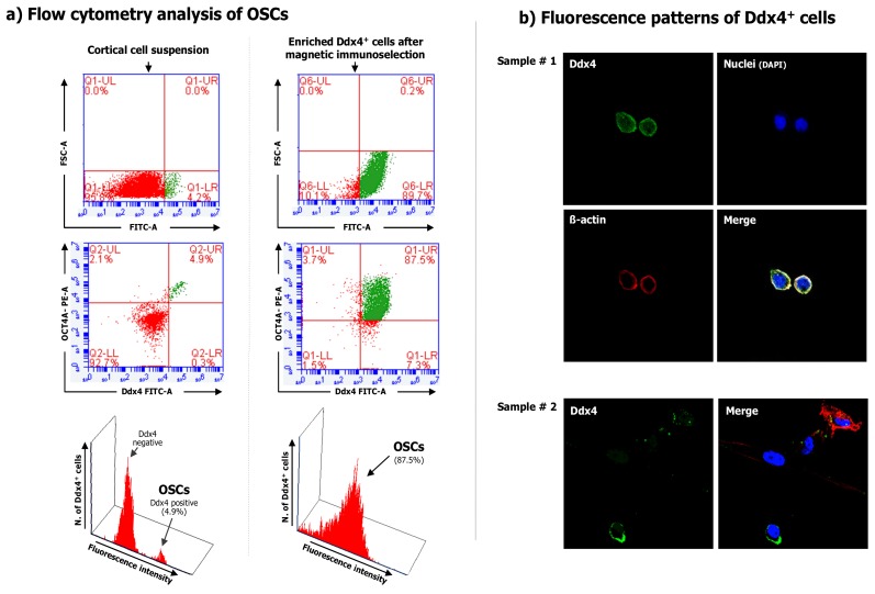
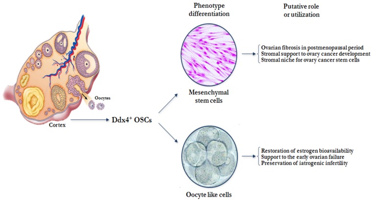
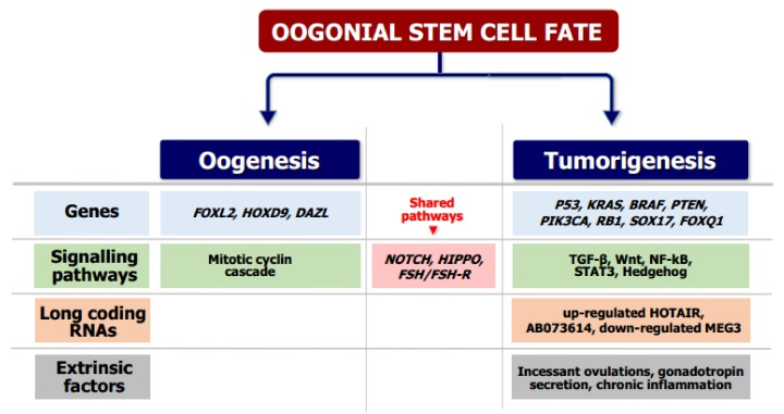
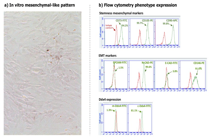
Recent studies support the existence of oogonial stem cells (OSCs) in the ovarian cortex of different mammals, including women.These cells are characterized by small size, membrane expression of DEAD(Asp-Glu-Ala-Asp)-box polypeptide-4 (Ddx4), and stemness properties (such as self-renewal and clonal expansion) as well as the ability to differentiate in vitro into oocyte-like cells. However, the discovery of OSCs contrasts with the popular theory that there is a numerically defined oocyte pool for female fertility which undergoes exhaustion with menopause. Indeed, in the ovarian cortex of postmenopausal women OSCs have been detected that possess both viability and capability to differentiate into oocytes, which is similar to those observed in younger patients. The pathophysiological role of this cell population in aged women is still debated since OSCs, under appropriate stimuli, differentiate into somatic cells, and the occurrence of Ddx4+ cells in ovarian tumor samples also suggests their potential involvement in carcinogenesis. Although further investigation into these observations is needed to clarify OSC function in ovary physiology, clinical investigators and researchers studying female infertility are presently focusing on OSCs as a novel opportunity to restore ovarian reserve in both young women undergoing early ovarian failure and cancer survivors experiencing iatrogenic menopause.

Keywords: Ddx4; infertility; menopause; oogonial stem cells; ovarian failure.


Conflict of interest statement

The authors declare no conflicts of interest.

Figures

Figure 1
Figure 1
Ddx4 expression on the cell population purified from the ovarian cortex.(a) Flow cytometry for Ddx4 expression measured in both total cortical cell suspension (left) and after Ddx4+ cell selection emphasized the small population extent in the cortex (4.9%) and its subsequent enrichment (87.5%). (b) Immunofluorescence for Ddx4 expression in two samples of purified Ddx4+ cells by confocal microscopy. The fluorescence patterns confirm the membrane localization of Ddx4 (FITC; green), while the cell integrity was assessed by both actin (phalloidin; red), and nuclei (DAPI; blue) staining.
Figure 2
Figure 2
Ddx4+ oogonial stem cells (OSCs). Representative image depicts the oocyte-like differentiation of OSCs (black arrows) after 21 days of culture in the presence of fibroblasts feeder layer. Although present in variable size during their differentiation, the largest cells (diameter of approximately 80–90 µm) acquired oocyte-like features, with prominent nuclei and perinuclear accumulation of organelles. By contrast, smaller cells were considered immature OSCs and might represent the oogonial stem cell reserve.
Figure 3
Figure 3
Hypothetical fate of Ddx4+ OSCs in post-menopausal women. Under the influence of different hormonal and micro environmental factors related to age or concurrent diseases, Ddx4+ OSCs might differentiate into mesenchymal stem cells and/or oocyte-like cells. In postmenopausal women, their mesenchymal differentiation could theoretically support ovarian fibrosis or stromal contribution to cancer development, whereas their oocyte-like induced differentiation could be utilized to restore ovarian hormonal failure or fertility.
Figure 4
Figure 4
Molecular signals involved in OSC differentiation. OSCs are able to undergo either neo-oogenesis or ovarian tumorigenesis depending on both different stimuli and activated pathways that in some cases are also shared. The molecular signals shown include well-established mechanisms and putative pathways, as reported in the literature [20,25,30,33].
Figure 5
Figure 5
In vitro mesenchymal differentiation and phenotype analysis of cultured Ddx4+ cells. (a) Light microscope image of 15-day cultured Ddx4+ cells in FSH and EGF-supplemented medium showing morphological variation from round to “spindle-like” adherent cells, with a typical aspect of fibroblasts or mesenchymal stem cells. (b) Flow cytometry analysis of cultured cells showed a mesenchymal phenotype with typical stemness markers (up), also including the epithelial-to-mesenchymal transition (EMT) molecules as N-cadherin (N-CAD) and CD146, whereas no epithelial markers as EPCAM and E-cadherin, were detected (middle). Based on their differentiation to the mesenchymal phenotype, OSCs lost Ddx4 membrane expression while the molecule was detectable in cytoplasm (down). Red histograms represent isotype controls used in flow cytometry assays.

References

    1. Zuckerman S. The number of oocytes in the mature ovary. Recent Prog. Horm. Res. 1951;6:63–108.
    1. Stimpfel M., Skutella T., Cvjeticanin B., Meznaric M., Dovc P., Novakovic S., Cerkovnik P., Vrtacnik-Bokal E., Virant-Klun I. Isolation, characterization and differentiation of cells expressing pluripotent/multipotent markers from adult human ovaries. Cell Tissue Res. 2013;354:593–607. doi: 10.1007/s00441-013-1677-8. - DOI - PubMed
    1. Zou K., Yuan Z., Yang Z., Luo H., Sun K., Zhou L., Xiang J., Shi L., Yu Q., Zhang Y., et al. Production of offspring from a germline stem cell line derived from neonatal ovaries. Nat. Cell Biol. 2009;11:631–636. doi: 10.1038/ncb1869. - DOI - PubMed
    1. Niikura Y., Niikura T., Tilly J.L. Aged mouse ovaries possess rare premeiotic germ cells that can generate oocytes following transplantation into a young host environment. Aging. 2009;1:971–978. doi: 10.18632/aging.100105. - DOI - PMC - PubMed
    1. Zhang D., Fouad H., Zoma W.D., Salama S.A., Wentz M.J., Al-Hendy A. Expression of stem and germ cell markers within nonfollicle structures in adult mouse ovary. Reprod. Sci. 2008;15:139–146. doi: 10.1177/1933719107310708. - DOI - PubMed

Publication types

Substances

LinkOut - more resources