. 2019 Jun 29;20(13):3191.
doi: 10.3390/ijms20133191.

Cystathionine-Gamma-Lyase-Derived Hydrogen Sulfide-Regulated Substance P Modulates Liver Sieve Fenestrations in Caecal Ligation and Puncture-Induced Sepsis

Affiliations

Cystathionine-Gamma-Lyase-Derived Hydrogen Sulfide-Regulated Substance P Modulates Liver Sieve Fenestrations in Caecal Ligation and Puncture-Induced Sepsis

Ravinder R Gaddam et al. Int J Mol Sci. .

Abstract

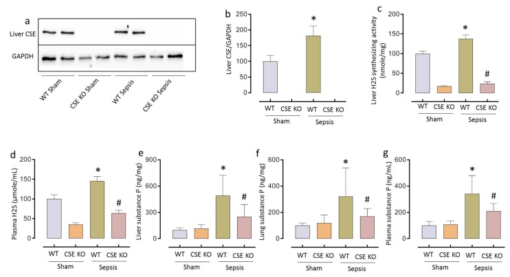
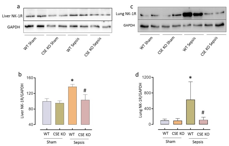
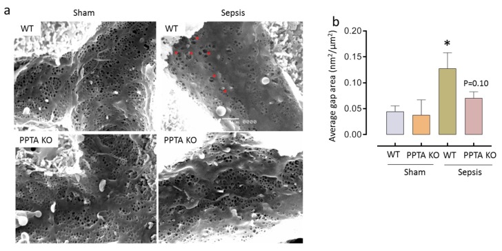
Cystathionine-γ-lyase (CSE) isa hydrogen sulfide (H2S)-synthesizing enzyme that promotesinflammation by upregulating H2S in sepsis. Liver sinusoidal endothelial cells (LSECs) are fenestrated endothelial cells (liver sieve) that undergo alteration during sepsis and H2S plays a role in this process. Substance P (SP) is encoded by the preprotachykinin A (PPTA) gene, and promotes inflammation in sepsis; however, its regulation by H2S is poorly understood. Furthermore, the interaction between H2S and SP in modulating LSEC fenestrations following sepsis remains unclear. This study aimed to investigate whether CSE/H2S regulates SP and the neurokinin-1 receptor (NK-1R) andmodulates fenestrations in LSECs following caecalligation and puncture (CLP)-induced sepsis. Here we report thatthe absence of either CSE or H2S protects against liver sieve defenestration and gaps formation in LSECsin sepsis by decreased SP-NK-1R signaling. Following sepsis, there is an increased expression of liver CSE and H2S synthesis, and plasma H2S levels, which were aligned with higher SP levels in the liver, lungs and plasma and NK-1R in the liver and lungs. The genetic deletion of CSE led to decreased sepsis-induced SP and NK-1R in the liver, lungs and plasma SP suggesting H2S synthesized through CSE regulates the SP-NK-1R pathway in sepsis. Further, mice deficient in the SP-encoding gene (PPTA) preservedsepsis-induced LSEC defenestrationand gaps formation, as seen by maintenance of patent fenestrations and fewer gaps. In conclusion, CSE/H2S regulates SP-NK-1R and modulates LSEC fenestrations in sepsis.

Keywords: cystathionine-gamma-lyase; hydrogen sulfide; liver sieve; neurokinin-1 receptor; substance P.


Conflict of interest statement

The authors declare no conflict of interest. The funders had no role in the design of the study, collection, analysis, and interpretations of the data, writing of the manuscript and in the decision to publish the results.

Figures

Figure 1
Figure 1
CSE-derived H2S regulates SP following CLP-induced sepsis. (ad) Increased liver CSE expression (a) and quantification of CSE in ‘a’ (b), liver H2S-synthesizing activity (c) and plasma H2S levels (d) following CLP-induced sepsis. (e–g) Genetic deletion of CSE significantly decreased SP levels in liver (e), lung (f) and plasma (g) compared to WT mice following sepsis. Data represented as mean ± S.E.M. (n = 8). The significance of differences among groups was evaluated by ANOVA with post-hoc Tukey’s test. Statistical significance was assigned as p < 0.05 (* p < 0.05 vs. WT sham; # p < 0.05 vs. WT sepsis).
Figure 2
Figure 2
CSE-derived H2S regulates NK-1R following CLP-induced sepsis. (ad) Genetic deletion of CSE significantly reduced liver (a) and lung (c) NK-1R protein expression compared to WT mice following sepsis and quantification of liver (b) and lung (d) NK-1R protein expression in ‘a’ and ‘c’, respectively. Data represented as mean ± S.E.M. (n = 8). The significance of differences among groups was evaluated by ANOVA with post-hoc Tukey’s test. Statistical significance was assigned as p < 0.05 (* p < 0.05 vs. WT sham; # p < 0.05 vs. WT sepsis).
Figure 3
Figure 3
PPTA KO mice protect against sepsis-induced LSEC defenestration and gaps formation. (a,b) Representative images of liver sieve fenestration micrographs (a) and average gap area of liver sieve fenestrae (b). LSEC injury significantly increased (as evidenced by gaps formation) following CLP-induced sepsis in WT mice. Genetic deletion of PPTA in mice showed fewer gaps following sepsis compared to WT sepsis mice. Data represented as mean ± S.E.M. (n = 4). The significance of differences among groups was evaluated by ANOVA with post-hoc Tukey’s test. Statistical significance was assigned as p < 0.05 (* p < 0.05 vs. WT sham; # p < 0.05 vs. WT sepsis).

References

    1. Zhao K., Li H., Li S., Yang G. Regulation of cystathionine gamma-lyase/H₂S system and its pathological implication. Front. Biosci. (Landmark edition) 2013;19:1355–1369. doi: 10.2741/4286. - DOI - PubMed
    1. Zhang H., Zhi L., Moochhala S.M., Moore P.K., Bhatia M. Endogenous hydrogen sulfide regulates leukocyte trafficking in cecal ligation and puncture-induced sepsis. J. Leukoc. Biol. 2007;82:894–905. doi: 10.1189/jlb.0407237. - DOI - PubMed
    1. Zhang H., Zhi L., Moore P.K., Bhatia M. Role of hydrogen sulfide in cecal ligation and puncture-induced sepsis in the mouse. Am. J. Physiol. Lung Cell. Mol. Physiol. 2006;290:L1193–L1201. doi: 10.1152/ajplung.00489.2005. - DOI - PubMed
    1. Badiei A., Chambers S., Gaddam R., Bhatia M. Cystathionine-γ-lyase gene silencing with siRNA in monocytes/macrophages attenuates inflammation in cecal ligation and puncture-induced sepsis in the mouse. J. Biosci. 2016;41:87–95. doi: 10.1007/s12038-016-9598-9. - DOI - PubMed
    1. Zhang H., Moochhala S.M., Bhatia M. Endogenous hydrogen sulfide regulates inflammatory response by activating the ERK pathway in polymicrobial sepsis. J. Immunol. 2008;181:4320–4331. doi: 10.4049/jimmunol.181.6.4320. - DOI - PubMed