. 2019 Jun 29;20(13):3197.
doi: 10.3390/ijms20133197.

Telmisartan Inhibits Cell Proliferation and Tumor Growth of Esophageal Squamous Cell Carcinoma by Inducing S-Phase Arrest In Vitro and In Vivo

Affiliations

Telmisartan Inhibits Cell Proliferation and Tumor Growth of Esophageal Squamous Cell Carcinoma by Inducing S-Phase Arrest In Vitro and In Vivo

Takanori Matsui et al. Int J Mol Sci. .

Abstract

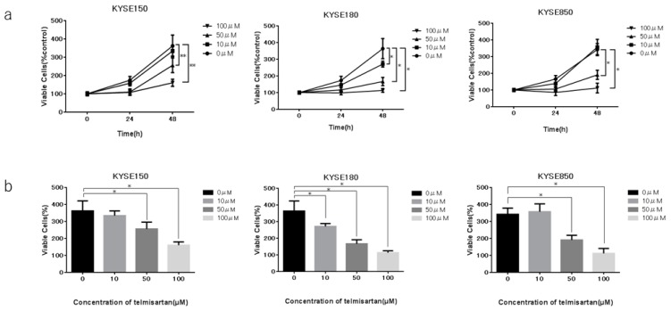
Esophageal squamous cell carcinoma (ESCC) is the most common primary esophageal malignancy. Telmisartan, an angiotensin II type 1 (AT1) receptor blocker (ARB) and a widely used antihypertensive, has been shown to inhibit proliferation of various cancer types. This study evaluated the effects of telmisartan on human ESCC cell proliferation in vitro and in vivo and sought to identify the microRNAs (miRNAs) involved in these antitumor effects. We examined the effects of telmisartan on three human ESCC cell lines (KYSE150, KYSE180, and KYSE850). Telmisartan inhibited proliferation of these three cell lines by inducing S-phase arrest, which was accompanied by decreased expression of cyclin A2, cyclin-dependent kinase 2, and other cell cycle-related proteins. Additionally, telmisartan reduced levels of phosphorylated ErbB3 and thrombospondin-1 in KYSE180 cells. Furthermore, expression of miRNAs was remarkably altered by telmisartan in vitro. Telmisartan also inhibited tumor growth in vivo in a xenograft mouse model. In conclusion, telmisartan inhibited cell proliferation and tumor growth in ESCC cells by inducing cell-cycle arrest.

Keywords: angiotensin II type 1 receptor blocker; cell cycle arrest; cyclin; esophageal squamous cell carcinoma; telmisartan.


Conflict of interest statement

The authors declare no conflict of interest.

Figures

Figure 1
Figure 1
Effects of telmisartan on proliferation of ESCC cell lines in vitro. Telmisartan inhibited proliferation of ESCC cells. Viability of treated cells differed significantly from that of control cells. (a) KYSE150, KYSE180, and KYSE850 cells were seeded in 96-well plate (1.0 × 10⁴ cells/well). After 24 h, telmisartan (10, 50, or 100 µM) or DMSO were added to the fresh culture medium. Cell viability was assayed daily from 0 to 48 h. (b) Cell viability of ESCC cells at 48 h. (*p < 0.01).
Figure 2
Figure 2
Flow cytometric analysis of KYSE180 cells treated with 50 µM telmisartan at 24 h. Telmisartan increased the population of cells in the S phase and decreased the population of cells in the G2/M phase. Telmisartan blocks cell-cycle progression to G2/M from S phase. (*p < 0.01).
Figure 3
Figure 3
Western blot analysis of cell-cycle regulatory proteins in KYSE180 cells treated with 50 µM telmisartan. Expression levels of Cyclin A2, Cyclin B1, CDK1, CDK2, CDK4 were decreased in treated cells.
Figure 4
Figure 4
Telmisartan does not promote apoptosis in KYSE180 cells. Flow cytometry assessment of apoptosis of KYSE180 cells treated with 50 µM telmisartan at 24 h. Percentages of Annexin V+ cells did not significantly differ between control cells and telmisartan-treated cells. Apoptosis in KYSE180 is not induced by telmisartan.
Figure 5
Figure 5
Result of p-RTK array for KYSE180 cells. (a) Template shows locations of tyrosine kinase antibodies on human p-RTK array. (b) p-ErbB3 expression was decreased in KYSE180 cells treated with 50 µM telmisartan at 24 h. (c) Densitometric ratio of telmisartan-treated group to non-treated group for p-ErbB3 spots. * p < 0.01.
Figure 6
Figure 6
Angiogenesis antibody array in KYSE180 cells. (a) Template shows locations of angiogenesis antibodies on human angiogenesis array. (b) TSP-1 expression was decreased in KYSE180 cells treated with 50 µM telmisartan at 24 h. (c) Densitometric ratio of telmisartan-treated group to the non-treated group for TSP-1 spots. * p < 0.01.
Figure 7
Figure 7
Hierarchical clustering of KYSE180 cells treated with or without telmisartan. KYSE180 cells were clustered according to expression profiles of 60 miRNAs differentially expressed by KYSE180 cells, with or without telmisartan. Top: miRNA clustering color scale indicates relative miRNA expression levels; red: high expression, blue: low expression.
Figure 8
Figure 8
KYSE180 tumors in nude mice treated with telmisartan. (a) Representative images of gross KYSE180 tumors treated with telmisartan (10 µg/day or 50 µg/day), or the control. (b) Growth curves of xenografts showing mean tumor volume of control and telmisartan at 10 µg/day or 50 µg/day). Tumors were significantly smaller in the high-dose (50 µg/day) group than in the control group. (c) Immunohistochemistry examination using PCNA staining (400× magnification). (d) Quantitative comparison of PCNA+ cells of the control, low-dose (10 µg/day) and high-dose (50 µg/day) groups. PCNA+ cells and total numbers of cells were counted from 5 fields (400× magnification). Percentages of PCNA+ cells were significantly smaller in low-dose and high-dose telmisartan treated groups than in the control group. * p < 0.05; ** p < 0.01.

References

    1. Bray F., Ferlay J., Soerjomataram I., Siegel R.L., Torre L.A., Jemal A. Global cancer statistics 2018: GLOBOCAN estimates of incidence and mortality worldwide for 36 cancers in 185 countries. CA A Cancer J. Clin. 2018;68:394–424. doi: 10.3322/caac.21492. - DOI - PubMed
    1. World Cancer Research Fund International Oesophageal Cancer Report 2016. [(accessed on 10 June 2019)]; Available online: http://www.wcrf.org/dietandcancer/oesophageal-cancer.
    1. Smyth E.C., Lagergren J., Fitzgerald R.C., Lordick F., Shah M.A., Lagergren P. Cunningham, D. Oesophageal cancer. Nat. Rev. Dis. Prim. 2017;3:17048. doi: 10.1038/nrdp.2017.48. - DOI - PMC - PubMed
    1. Pennathur A., Gibson M.K., Jobe B.A., Luketich J.D. Oesophageal carcinoma. Lancet. 2013;381:400–412. doi: 10.1016/S0140-6736(12)60643-6. - DOI - PubMed
    1. Furihata M., Ishikawa T., Inoue A., Yoshikawa C., Sonobe H., Ohtsuki Y., Araki K., Ogoshi S. Determination of the prognostic significance of unscheduled cyclin A overexpression in patients with esophageal squamous cell carcinoma. Clin. Cancer Res. 1996;2:1781–1785. - PubMed

MeSH terms