The Antiviral Agent Cidofovir Induces DNA Damage and Mitotic Catastrophe in HPV-Positive and -Negative Head and Neck Squamous Cell Carcinomas In Vitro
- PMID: 31262012
- PMCID: PMC6678333
- DOI: 10.3390/cancers11070919
The Antiviral Agent Cidofovir Induces DNA Damage and Mitotic Catastrophe in HPV-Positive and -Negative Head and Neck Squamous Cell Carcinomas In Vitro
Abstract
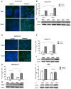
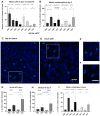
Cidofovir (CDV) is an antiviral agent with antiproliferative properties. The aim of our study was to investigate the efficacy of CDV in HPV-positive and -negative head and neck squamous cell carcinoma (HNSCC) cell lines and whether it is caused by a difference in response to DNA damage. Upon CDV treatment of HNSCC and normal oral keratinocyte cell lines, we carried out MTT analysis (cell viability), flow cytometry (cell cycle analysis), (immuno) fluorescence and western blotting (DNA double strand breaks, DNA damage response, apoptosis and mitotic catastrophe). The growth of the cell lines was inhibited by CDV treatment and resulted in γ-H2AX accumulation and upregulation of DNA repair proteins. CDV did not activate apoptosis but induced S- and G2/M phase arrest. Phospho-Aurora Kinase immunostaining showed a decrease in the amount of mitoses but an increase in aberrant mitoses suggesting mitotic catastrophe. In conclusion, CDV inhibits cell growth in HPV-positive and -negative HNSCC cell lines and was more profound in the HPV-positive cell lines. CDV treated cells show accumulation of DNA DSBs and DNA damage response activation, but apoptosis does not seem to occur. Rather our data indicate the occurrence of mitotic catastrophe.
Keywords: Aurora Kinase A; DNA repair; cell line; cyclin B1; double-stranded DNA breaks; head and neck cancer; human papillomavirus.
Conflict of interest statement
Ernst-Jan Speel has received research grants from Pfizer and Novartis. The other authors disclose no potential conflicts of interest.
Figures


Similar articles
-
Cidofovir is active against human papillomavirus positive and negative head and neck and cervical tumor cells by causing DNA damage as one of its working mechanisms.Oncotarget. 2016 Jul 26;7(30):47302-47318. doi: 10.18632/oncotarget.10100. Oncotarget. 2016. PMID: 27331622 Free PMC article.
-
Cidofovir selectivity is based on the different response of normal and cancer cells to DNA damage.BMC Med Genomics. 2013 May 23;6:18. doi: 10.1186/1755-8794-6-18. BMC Med Genomics. 2013. PMID: 23702334 Free PMC article.
-
Synthetic Lethal Targeting of Mitotic Checkpoints in HPV-Negative Head and Neck Cancer.Cancers (Basel). 2020 Jan 28;12(2):306. doi: 10.3390/cancers12020306. Cancers (Basel). 2020. PMID: 32012873 Free PMC article.
-
Induction of apoptosis by cidofovir in human papillomavirus (HPV)-positive cells.Oncol Res. 2000;12(9-10):397-408. doi: 10.3727/096504001108747855. Oncol Res. 2000. PMID: 11697818
-
Cidofovir in the treatment of HPV-associated lesions.Verh K Acad Geneeskd Belg. 2001;63(2):93-120, discussion 120-2. Verh K Acad Geneeskd Belg. 2001. PMID: 11436421 Review.
Cited by
-
SEC11A contributes to tumour progression of head and neck squamous cell carcinoma.Heliyon. 2023 Mar 28;9(4):e14958. doi: 10.1016/j.heliyon.2023.e14958. eCollection 2023 Apr. Heliyon. 2023. PMID: 37025806 Free PMC article.
-
Revealing the viral culprits: the hidden role of the oral virome in head and neck cancers.Arch Microbiol. 2025 Feb 28;207(4):73. doi: 10.1007/s00203-025-04270-x. Arch Microbiol. 2025. PMID: 40095096 Free PMC article. Review.
-
Human Papillomavirus and Cancers.Cancers (Basel). 2020 Dec 15;12(12):3772. doi: 10.3390/cancers12123772. Cancers (Basel). 2020. PMID: 33333750 Free PMC article.
-
Presence of Human Papillomavirus and Epstein-Barr Virus, but Absence of Merkel Cell Polyomavirus, in Head and Neck Cancer of Non-Smokers and Non-Drinkers.Front Oncol. 2021 Jan 20;10:560434. doi: 10.3389/fonc.2020.560434. eCollection 2020. Front Oncol. 2021. PMID: 33552950 Free PMC article.
-
Newly Synthesized Melphalan Analogs Induce DNA Damage and Mitotic Catastrophe in Hematological Malignant Cancer Cells.Int J Mol Sci. 2022 Nov 17;23(22):14258. doi: 10.3390/ijms232214258. Int J Mol Sci. 2022. PMID: 36430734 Free PMC article.
References
LinkOut - more resources
Full Text Sources

