. 2019 Jul 1;20(1):117.
doi: 10.1186/s12881-019-0854-3.

Overexpression of MicroRNA-148b-3p stimulates osteogenesis of human bone marrow-derived mesenchymal stem cells: the role of MicroRNA-148b-3p in osteogenesis

Affiliations

Overexpression of MicroRNA-148b-3p stimulates osteogenesis of human bone marrow-derived mesenchymal stem cells: the role of MicroRNA-148b-3p in osteogenesis

Samaneh Mollazadeh et al. BMC Med Genet. .

Abstract

Background: Mesenchymal stem cells (MSCs) are attractive choices in regenerative medicine and can be genetically modified to obtain better results in therapeutics. Bone development and metabolism are controlled by various factors including microRNAs (miRs) interference, which are small non-coding endogenous RNAs.

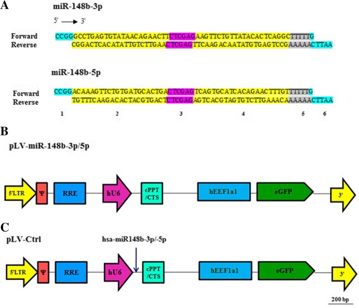
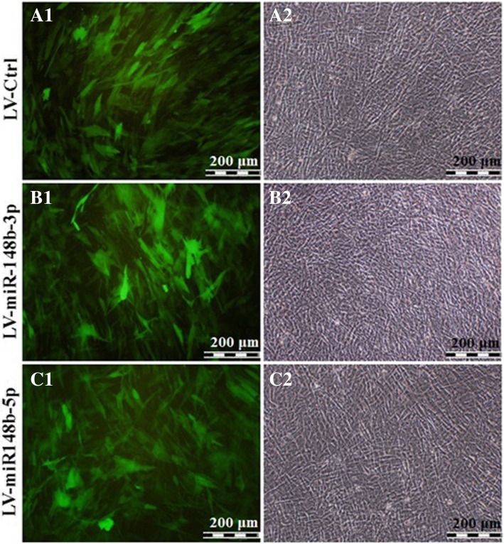
Methods: In the current study, the effects of forced miR-148b expression was evaluated on osteogenic activity. Human bone marrow-derived mesenchymal stem cells (BM-MSCs) were transduced with bicistronic lentiviral vector encoding hsa-miR-148b-3p or -5p and the enhanced green fluorescent protein. Fourteen days post-transduction, immunostaining as well as Western blotting were used to analyze osteogenesis.

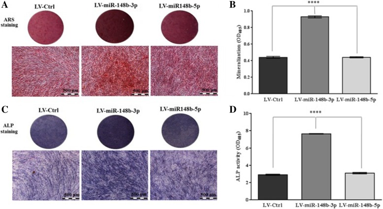
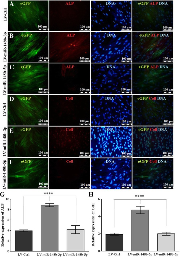
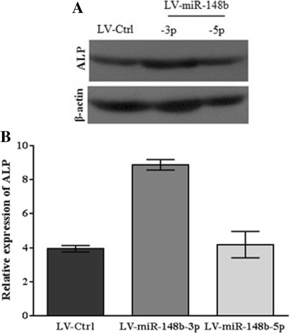
Results: Overexpression of miR-148b-3p increased the osteogenic differentiation of human BM-MSCs as demonstrated by anenhancement of mineralized nodular formation and an increase in the levels of osteoblastic differentiation biomarkers, alkaline phosphatase and collagen type I.

Conclusions: Since lentivirally overexpressed miR-148b-3p increased osteogenic differentiation capability of BM-MSCs, this miR could be applied as a therapeutic modulator to optimize bone function.

Keywords: Lentivirus; Mesenchymal stem cells; MicroRNA-148b; Osteogenesis.


Conflict of interest statement

The authors declare that they have no competing interest.

Figures

Fig. 1
Fig. 1
Cassettes and structure of LV shuttle plasmids. a Schematic representation of miR-148b-3p and miR-148b-5p sequences after ligation of forward and reverse primers. Section 1 and 6 are SgrA1 and EcoRI restriction sites, respectively. Section 2 and 4 are sense and antisense strands, respectively. Section 3 is necessary for hairpin structure (loop). Section 5 is a terminator (poly T). b Maps of pLV-miR-148b-3p and pLV-miR-148b-5p (c) and of pLV-Ctrl.5′ LTR, chimeric 5′ long terminal repeat including the Rous sarcoma virus U3 region as well as the HIV1 R and U5 regions; Ψ, HIV1 packaging signal; RRE, HIV1 Rev-responsive element; hU6, human U6 gene promoter; miR-158b-3p/5p, coding sequence of desired miRNAs; cPPT/CTS, HIV1 central polypurine tract and termination site; hEEF1a1, eukaryotic translation elongation factor 1 alpha 1 gene promoter; EGFP; enhanced green fluorescent coding sequence; 3′ LTR, 3′ HIV1 long terminal repeat involving a deletion in the U3 region to render the LV self-inactivating
Fig. 2
Fig. 2
Fluoromicrographs (A1, B1, C1), and bright field (A2, B2, C2) images of LV-Ctrl, LV-miR-148b-3p, and LV-miR-148b-5p-transduced human BM-MSCs that were cultured in osteogenic differentiation medium for 21 days
Fig. 3
Fig. 3
Increased expression of miR-148b-3p in human BM-MSCs was potentially mediated by LV enhanced osteogenic differentiation. a After 21 days mineral nodules depositions were detected with ARS staining. b The stained cells were eluted using 10% acetic acid and the absorbance of extracted stain was measured at 405 nm to quantify the mineralization level. c ALP production was evaluated with BCIP/NBT kit as dark purples deposits. d After 14 days ALP activity was quantified with pNPP substrate system.****P < 0.0001
Fig. 4
Fig. 4
Osteogenic differentiation of human BM-MSCs was increased by miR-148b-3p overexpression. (a-f) Representative micrograph of 14-day incubation of human BM-MSCs exposed to LV-Ctrl, LV-miR-148b-3p or LV-miR-148b-5p. Immunostaining of ALP and ColI with nuclear counterstained (Hoechst) were shown in red and blue respectively. It should be mentioned that EGFP+ cells were in green. (g-h)BM-MSCs overexpressed miR-148b-3p demonstrated 2.2- and 2.5-fold enhancement in ALP and ColI expression respectively compared to other groups. ****shows significant differences (P < 0.0001) between LV-miR-148b-3pwith LV-miR-148b-5p and LV-Ctrl
Fig. 5
Fig. 5
The exogenous overexpression of miR-148b-3p upregulated protein expression level of ALP. a 14 days post-transduction, ALP were expressed by the cells in all infected groups (LV-Ctrl, LV-miR-148b-3p, LV-miR-148b-5p). In contrast, ALP exhibited greater expressionin the cells treated with miR-148b-3p. The resultswere shown by the band density ratio of ALP with β-actin. b The relative expression of ALP (fold change) after quantification of Western blotting analysis

References

    1. Abdallah B, Kassem M. Human mesenchymal stem cells: from basic biology to clinical applications. Gene Ther. 2008;15(2):1092. - PubMed
    1. Kassem M, Abdallah BM. Human bone-marrow-derived mesenchymal stem cells: biological characteristics and potential role in therapy of degenerative diseases. Cell Tissue Res. 2008;331(1):157–163. - PubMed
    1. Zhang W-B, Zhong W-J, Wang L. A signal-amplification circuit between miR-218 and Wnt/β-catenin signal promotes human adipose tissue-derived stem cells osteogenic differentiation. Bone. 2014;58:59–66. - PubMed
    1. Qureshi AT, Monroe WT, Dasa V, Gimble JM, Hayes DJ. miR-148b–nanoparticle conjugates for light mediated osteogenesis of human adipose stromal/stem cells. Biomaterials. 2013;34(31):7799–7810. - PubMed
    1. Tiago DM, Marques CL, Roberto VP, Cancela ML, Laizé V. Mir-20a regulates in vitro mineralization and BMP signaling pathway by targeting BMP-2 transcript in fish. Arch Biochem Biophys. 2014;543:23–30. - PubMed

Publication types