. 2019 Jul;21(7):867-878.
doi: 10.1038/s41556-019-0348-8. Epub 2019 Jul 1.

Phosphorylated Rho-GDP directly activates mTORC2 kinase towards AKT through dimerization with Ras-GTP to regulate cell migration

Affiliations

Phosphorylated Rho-GDP directly activates mTORC2 kinase towards AKT through dimerization with Ras-GTP to regulate cell migration

Hiroshi Senoo et al. Nat Cell Biol. 2019 Jul.

Abstract

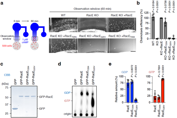
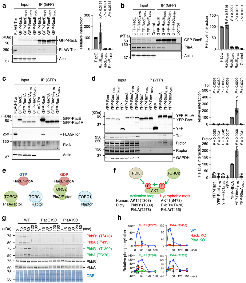
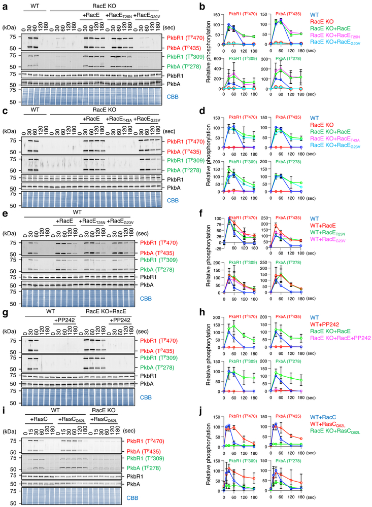
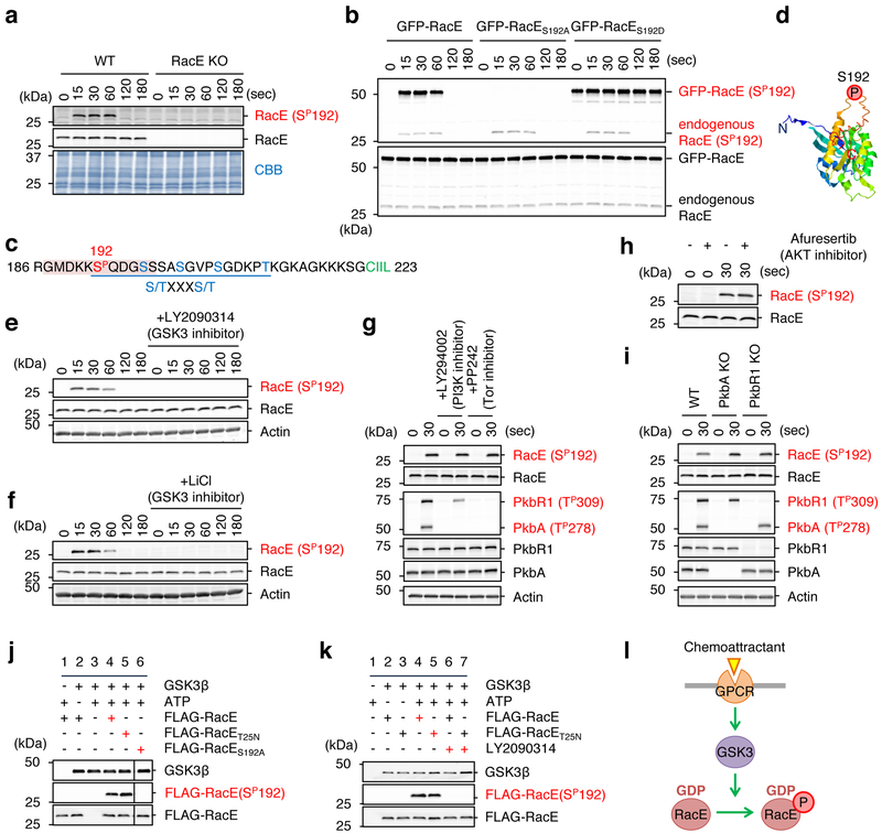
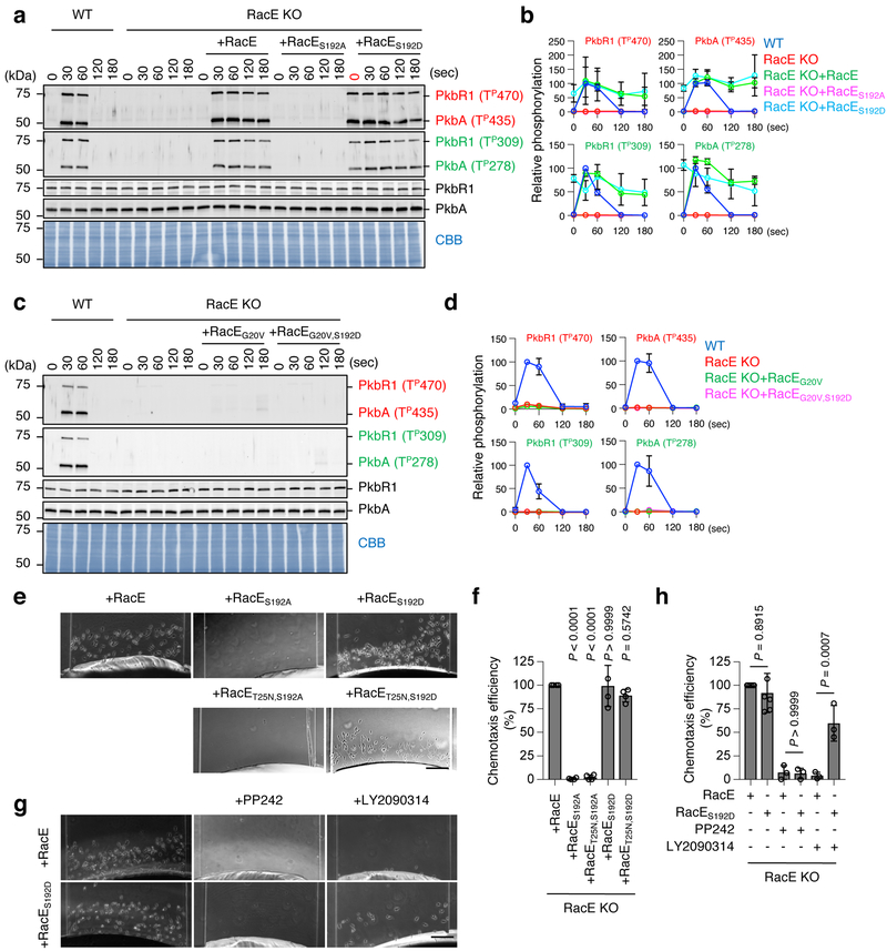
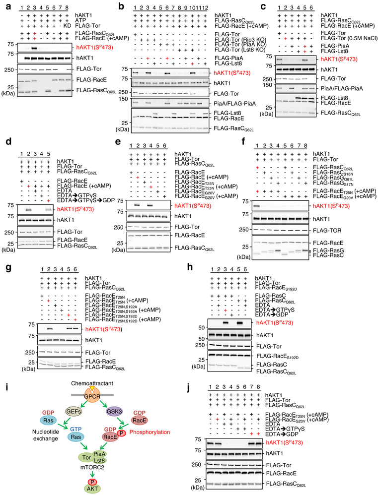
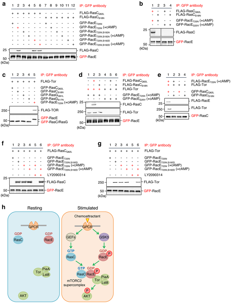
mTORC2 plays critical roles in metabolism, cell survival and actin cytoskeletal dynamics through the phosphorylation of AKT. Despite its importance to biology and medicine, it is unclear how mTORC2-mediated AKT phosphorylation is controlled. Here, we identify an unforeseen principle by which a GDP-bound form of the conserved small G protein Rho GTPase directly activates mTORC2 in AKT phosphorylation in social amoebae (Dictyostelium discoideum) cells. Using biochemical reconstitution with purified proteins, we demonstrate that Rho-GDP promotes AKT phosphorylation by assembling a supercomplex with Ras-GTP and mTORC2. This supercomplex formation is controlled by the chemoattractant-induced phosphorylation of Rho-GDP at S192 by GSK-3. Furthermore, Rho-GDP rescues defects in both mTORC2-mediated AKT phosphorylation and directed cell migration in Rho-null cells in a manner dependent on phosphorylation of S192. Thus, in contrast to the prevailing view that the GDP-bound forms of G proteins are inactive, our study reveals that mTORC2-AKT signalling is activated by Rho-GDP.


Figures

Figure 1.
Figure 1.. RacE-GDP functions in directed cell migration.
a-b, Cell migration toward the chemoattractant cAMP was analyzed in WT and RacE-KO Dictyostelium cells carrying WT RacE, GDP-bound RacET25N, GTP-bound RacEG20V or effector domain-defective RacET43A for 60 min in a microfluidic chamber in a. Bar, 100 μm. Chemotaxis efficiency was quantified by measuring the number of cells that moved toward a higher concentration of cAMP (observation window) in b. The chemotaxis efficiency in RacE-KO cells expressing WT RacE (KO+RacE) was set 100% (n = 4 independent experiments). Values are means ± SD. Significance was calculated using ANOVA with post-hoc Tukey. p values are shown for comparison of KO+RacE with others. c-e, Guanine nucleotide binding to RacE. Differentiated Dictyostelium cells carrying GFP, GFP-RacE, GFP-RacET25N or GFP-RacEG20V were metabolically labeled using P for 1 h. GFP fusion proteins were immunopurified using GFP-Trap beads and analyzed using SDS-PAGE and CBB staining in c. Bound guanine nucleotides were analyzed by thin layer chromatography and phosphoimaging in d. Quantification of GDP and GTP (n=5 independent experiments for GFP-RacE and GFP-RacEG20V and n=4 independent experiments for GFP-RacET25N) in e. Values are means ± SD. Significance was calculated using ANOVA with post-hoc Tukey. p values are shown for comparison of GFP-RacE with others.
Figure 2.
Figure 2.. RacE-GDP specifically interacts with mTORC2.
a, Dictyostelium cell lysates carrying GFP fused to the indicated forms of RacE were incubated with cell lysates carrying FLAG-Tor and subjected to immunoprecipitation with GFP-Trap. Quantification of interaction is shown. The band intensity of FLAG-Tor in immunoprecipitates of cells expressing WT RacE was set 100% (n=6, 6, 6 and 3 independent experiments for RacE, RacET25N, RacEG20V and RacET43A, respectively). Values are average ± SD. Significance was calculated using ANOVA with post-hoc Tukey. p values are shown for comparison between RacE and others. b, Dictyostelium cell lysates carrying the indicated GFP-RacE were subject to immunoprecipitation with GFP-Trap to analyze its association with endogenous PiaA. The band intensity of PiaA in immunoprecipitates of cells expressing WT RacE was set 100% (n = 3 independent experiments). Values are average ± SD. Significance was calculated using ANOVA with post-hoc Tukey. p values are shown for comparison betwen RacE with others. c, Dictyostelium cell lysates carrying GFP fused to the indicated forms of Rac1A and RacE were incubated with cell lysates carrying FLAG-Tor and subjected to immunoprecipitation with GFP-Trap. Experiment was repeated independently three times with similar results. d, HEK293T cells were transfected with YFP fused to the indicated constructs of human Rac1 and RhoA and subjected to immunoprecipitation using GFP-Trap. The band intensities of Tor and rictor in immunoprecipitates of cells expressing WT RhoA was set 100% (n = 3 and n = 4 independent experiments for Tor and Rictor, respectively). Values are average ± SD. Significance was calculated using ANOVA with post-hoc Tukey. p values are shown for comparison between YFP-RhoA and others. e, Summary of the data. f, AKTs are phosphorylated in the hydrophobic motif by mTORC2 and in the activation loop by PDK. g and h, WT, RacE-KO, and PiaA-KO Dictyostelium cells were stimulated with the chemoattractant cAMP (1 μM). (n = 3 independent experiments). The total amounts of two AKT homologs (PkbR1 and PkbA) and their phosphorylation (Red: hydrophobic motif, Green: activation loop) were analyzed by immunoblotting. PVDF membranes were stained with CBB as loading controls. The band intensity of phosphorylated AKTs was quantified in h: WT cells at 30 s were set at 100%. Values are average ± SD.
Figure 3.
Figure 3.. RacE-GDP promotes chemoattractant-induced, mTORC2-mediated AKT phosphorylation in cells.
The indicated Dictyostelium cell lines were stimulated with the chemoattractant cAMP (1 μM). a-f, WT cells and RacE-KO cells expressing different GFP-RacE constructs were analyzed. g and h, WT cells and RacE-KO cells expressing GFP-RacE were pretreated with 0.5 μM of the mTORC2 inhibitor PP242 for 10 min and then stimulated with cAMP. i and j, WT and RacE-KO cells expressing FLAG-tagged RasC or GTP-bound RasCQ62L were analyzed. a-j, Total amounts of two AKT homologs (PKBR1 and PKBA) and their phosphorylation (Red: hydrophobic motif, Green: activation loop) were analyzed by immunoblotting. PVDF membranes were stained with CBB as loading controls in a, c, e, g, and i. The band intensity of phosphorylated AKTs was quantified in b, d, f, h, and j: WT cells at 30 s (b, d, f and h) and WT cells expressing RasC at 30 s (j) were set at 100%. Values are average ± SD (n = 3 independent experiments).
Figure 4.
Figure 4.. GSK-3 phosphorylates RacE at Ser192 in response to the chemoattractant.
a, WT and RacE-KO cells were stimulated with the chemoattractant cAMP (1 μM). Total amounts of RacE and its phosphorylation at Ser192 were analyzed by immunoblotting with antibodies to RacE and phospho-RacE(Ser192). b, WT cells expressing GFP fused to WT RacE, phospho-defective RacES192A or phospho-mimetic RacES192D were stimulated with cAMP. Whole cell lysates prepared at the indicated time points were analyzed by immunoblotting with antibodies to RacE and phospho-RacE(S192). c, The amino acid sequence in the vicinity of the phosphorylation site (Ser192, red) of RacE. A consensus motif for GSK-3 phosphorylation — a cluster of (S/TXXXS/T) — is underlined. A phosphopeptide used to raise anti-phospho-RacE (S192P) is highlighted. d, The location of serine 192 in a modeled RacE 3-D structure. (e–h) WT cells were treated with inhibitors to GSK-3 (250 nM LY2090314 in e and 10 mM lithium in f), PI3K (250 μM LY294002 in g), mTORC2 (0.5 μM PP242 in g), or AKT (5 μM afuresertib in h) for 10 min. Cells were then stimulated by the chemoattractant cAMP for the indicated amounts of time. Whole cell lysates were analyzed by immunoblotting with antibodies to RacE and phospho-RacE(Ser192). i, WT and cells lacking AKT (PkbA-KO or PkbR1-KO) were stimulated with cAMP for 30 s. Whole cell lysates were analyzed by immunoblotting with the indicated antibodies. j, Purified human GSK-3β was incubated with purified WT, GDP-bound RacET25N or phospho-defective RacES192A for 15 min. Ser192 phosphorylation of RacE was tested by immunoblotting. k, WT or GDP-bound RacET25N was mixed with GSK-3β in the presence or absence of the GSK-3 inhibitor LY2090314 (10 nM) and examined for Ser192 phosphorylation using immunoblotting. l, Summary of the data. Experiments were repeated independently three times with similar results in a, b and e-k.
Figure 5.
Figure 5.. Ser192 phosphorylated RacE-GDP activates mTORC2.
a-d, WT cells and RacE-KO cells carrying different GFP-RacE constructs were stimulated by cAMP and analyzed for AKT phosphorylation. b and d, Quantification of band intensity (n = 3 independent experiments): WT cells at 30 s were set at 100%. e-h, Directed cell migration toward the chemoattractant cAMP was analyzed in RacE-KO cells carrying different GFP-RacE constructs in a microfluidic chamber in the presence or absence of inhibitors to mTORC2 (0.5 μM PP242) or GSK-3 (250 nM LY2090314). Bar, 100 μm. Chemotaxis efficiency was quantified in f and h for e and g, respectively. The chemotaxis efficiency in RacE-KO cells expressing WT RacE was set 100%. Values are average ± SD. Significance was calculated using ANOVA with post-hoc Tukey. p values are shown for comparison between RacE and others in f (n=4 independent experiments) and between untreated RacE and RacES129D, or between PP242-treated RacE and RacES129D, or LY2090314-treated RacE and RacES129D in f (n=6, 3, 3, 6, 3 and 3 independent experiments for untreated RacE, untreated RacES192D, PP242-treated RacE, PP242-treated RacES192D, LY2090314-treated RacE and LY2090314-treated RacES192D).
Figure 6.
Figure 6.. Phosphorylated RacE-GDP activates mTORC2 in vitro.
mTORC2-mediated AKT phosphorylation was reconstituted using purified proteins. mTORC2 (FLAG-Tor, -PiaA or -Lst8), FLAG-RacE, and FLAG-RasC/G were purified from Dictyostelium cells. +cAMP indicates that cells were stimulated by the chemoattractant for 30 s before purification of FLAG-RacE. Purified proteins were mixed in the presence or absence of ATP and human unactive AKT for 5 min at room temperature. AKT phosphorylation was analyzed by immunoblotting using anti-phospho AKT (serine 473) antibodies. a, mTORC2 phosphorylates AKT in the presence of RacE and RasC. b and c, mTORC2 activation requires PiaA and Lst8, but not the mSIN1 homolog Rip3. FLAG-PiaA or FLAG-Lst8 was added to FLAG-Tor purified from the indicated KO cell lines in b. FLAG-PiaA and/or FLAG-Lst8 were incubated with high-salt washed FLAG-Tor in c. d, mTORC2 activation requires RacE-GDP. Purified RacE was incubated with EDTA (25 mM), GTPγS (0.5 mM) or GTPγS then GDP (2.5 mM) (GTPγS → GDP) before reconstitution. e, WT RacE or GDP-bound RacET25N, but not GTP-bound RacEG20V, activates mTORC2 after the chemoattractant stimulation. f, mTORC2 activation needs RasC-GTP but not RasG-GTP. g, RacE phosphorylation controls mTORC2 activation. Phospho-mimetic mutation S192D in GDP-bound RacET25N activates mTORC2 without chemoattractant stimulation, while the phospho-defective S192A mutation blocks it. h, mTORC2 activation requires RasC-GTP. Purified RacC was incubated with EDTA followed by either GTPγS or GDP before reconstitution. i, Summary of the data. j, RacEG23V-GDP activates mTORC2. Purified RacET25N and RacEG23V were incubated with EDTA followed by GTPγS or GDP prior to reconstitution. Experiments were repeated independently three times with similar results in a-h and j.
Figure 7.
Figure 7.. Ser192 phosphorylated RacE-GDP forms a supercomplex with Tor and Ras-GTP.
a and b, The indicated GFP-RacE proteins were purified from Dictyostelium cells with or without 1 μM cAMP stimulation for 30 s. FLAG-RasC proteins were purified without cAMP stimulation. GFP-RacE was incubated with FLAG-RasC and pulled down using GFP-Trap. The pellet fraction was analyzed by immunoblotting using antibodies to GFP and FLAG. c, GFP-RacE, GFP-RasC or GFP-RasG was incubated with FLAG-Tor that was purified in a high-salt condition and pulled down with GFP-Trap. The pellet fraction was analyzed by immunoblotting using antibodies to GFP and FLAG. d, GFP fused to GDP-bound RacET25N or GTP-bound RacEG20V were purified from Dictyostelium cells under a high salt condition after stimulation with the chemoattractant cAMP. These GFP fusion proteins were incubated with high-salt washed FLAG-Tor and/or FLAG-RasC proteins. GFP-RacE was pulled down with GFP-Trap, and the pellet fractions were analyzed by immunoblotting. e, RacE forms a complex with Tor and RasC. The indicated proteins were purified under high-salt conditions and mixed for 15 min at room temperature. GFP-RasC proteins were pulled down with GFP-Trap, and the pellet fraction was analyzed by immunoblotting. f and g, Different GFP-RacE proteins were purified from Dictyostelium cells with or without 1 μM cAMP stimulation for 30 s in the presence or absence of the GSK-3 inhibitor LY2090314 (250 nM). GFP-RacE was incubated with FLAG-RasC in f or FLAG-Tor in g and pulled down using GFP-Trap. The pellet fraction was analyzed by immunoblotting using antibodies to GFP and FLAG. h, Model for GPCR-mediated mTORC2-AKT signaling. In response to GPCR activation by chemoattractant, Rho-GDP becomes phosphorylated by GSK-3 and assembles the super signaling complex with Ras-GTP and mTORC2 to promote AKT phosphorylation. Experiments were repeated independently three times with similar results in a-g.

Similar articles

Cited by

References

    1. Hodge RG & Ridley AJ Regulating Rho GTPases and their regulators. Nat Rev Mol Cell Biol 17, 496–510 (2016). - PubMed
    1. Burridge K & Wennerberg K Rho and Rac take center stage. Cell 116, 167–179 (2004). - PubMed
    1. Stenmark H Rab GTPases as coordinators of vesicle traffic. Nat Rev Mol Cell Biol 10, 513–525 (2009). - PubMed
    1. Alberts B et al. Molecular Biology of the Cell, Sixth Edition Molecular Biology of the Cell, Sixth Edition, 1–1342 (2015).
    1. Mayor R & Etienne-Manneville S The front and rear of collective cell migration. Nat Rev Mol Cell Biol 17, 97–109 (2016). - PubMed

Publication types

MeSH terms

Substances

LinkOut - more resources