

Modulation of Fronto-Striatal Functional Connectivity Using Transcranial Magnetic Stimulation
- PMID: 31263404
- PMCID: PMC6585467
- DOI: 10.3389/fnhum.2019.00190
Modulation of Fronto-Striatal Functional Connectivity Using Transcranial Magnetic Stimulation
Abstract
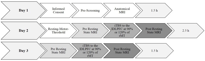
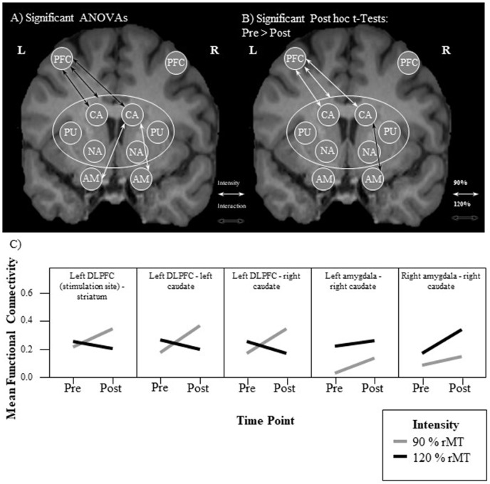
Background: The fronto-striatal network is involved in various motor, cognitive, and emotional processes, such as spatial attention, working memory, decision-making, and emotion regulation. Intermittent theta burst transcranial magnetic stimulation (iTBS) has been shown to modulate functional connectivity of brain networks. Long stimulation intervals, as well as high stimulation intensities are typically applied in transcranial magnetic stimulation (TMS) therapy for mood disorders. The role of stimulation intensity on network function and homeostasis has not been explored systematically yet. Objective: In this pilot study, we aimed to modulate fronto-striatal connectivity by applying iTBS at different intensities to the left dorso-lateral prefrontal cortex (DLPFC). We measured individual and group changes by comparing resting state functional magnetic resonance imaging (rsfMRI) both pre-iTBS and post-iTBS. Differential effects of individual sub- vs. supra-resting motor-threshold stimulation intensities were assessed. Methods: Sixteen healthy subjects underwent excitatory iTBS at two intensities [90% and 120% of individual resting motor threshold (rMT)] on separate days. Six-hundred pulses (2 s trains, 8 s pauses, duration of 3 min, 20 s) were applied over the left DLPFC. Directly before and 7 min after stimulation, task-free rsfMRI sessions, lasting 10 min each, were conducted. Individual seed-to-seed functional connectivity changes were calculated for 10 fronto-striatal and amygdala regions of interest with the SPM toolbox DPABI. Results: Sub-threshold-iTBS increased functional connectivity directly between the left DLPFC and the left and right caudate, respectively. Supra-threshold stimulation did not change fronto-striatal functional connectivity but increased functional connectivity between the right amygdala and the right caudate. Conclusion: A short iTBS protocol applied at sub-threshold intensities was not only sufficient, but favorable, in order to increase bilateral fronto-striatal functional connectivity, while minimizing side effects. The absence of an increase in functional connectivity after supra-threshold stimulation was possibly caused by network homeostatic effects.
Keywords: DLPFC; fronto-striatal network; functional connectivity; intermittent theta burst stimulation (iTBS); prefrontal cortex; resting state; striatum.
Figures
Similar articles
-
Increasing striatal dopamine release through repeated bouts of theta burst transcranial magnetic stimulation of the left dorsolateral prefrontal cortex. A 18F-desmethoxyfallypride positron emission tomography study.Front Neurosci. 2024 Jan 18;17:1295151. doi: 10.3389/fnins.2023.1295151. eCollection 2023. Front Neurosci. 2024. PMID: 38304075 Free PMC article.
-
Preconditioning prefrontal connectivity using transcranial direct current stimulation and transcranial magnetic stimulation.Front Hum Neurosci. 2022 Aug 11;16:929917. doi: 10.3389/fnhum.2022.929917. eCollection 2022. Front Hum Neurosci. 2022. PMID: 36034122 Free PMC article.
-
Dynamic Functional Connectivity Within the Fronto-Limbic Network Induced by Intermittent Theta-Burst Stimulation: A Pilot Study.Front Neurosci. 2019 Sep 13;13:944. doi: 10.3389/fnins.2019.00944. eCollection 2019. Front Neurosci. 2019. PMID: 31572111 Free PMC article.
-
Modulation of dorsolateral prefrontal cortex functional connectivity after intermittent theta-burst stimulation in depression: Combining findings from fNIRS and fMRI.Neuroimage Clin. 2022;34:103028. doi: 10.1016/j.nicl.2022.103028. Epub 2022 May 2. Neuroimage Clin. 2022. PMID: 35537216 Free PMC article. Clinical Trial.
-
The effects of intermittent theta burst stimulation (iTBS) on resting-state brain entropy (BEN).Neurotherapeutics. 2025 Apr;22(3):e00556. doi: 10.1016/j.neurot.2025.e00556. Epub 2025 Mar 5. Neurotherapeutics. 2025. PMID: 40050146 Free PMC article.
Cited by
-
Neural mechanisms of reward processing in preadolescent irritability: Insights from the ABCD study.J Affect Disord. 2025 Feb 1;370:286-298. doi: 10.1016/j.jad.2024.10.124. Epub 2024 Oct 31. J Affect Disord. 2025. PMID: 39488236 Free PMC article.
-
Repetitive Transcranial Magnetic Stimulation for Adolescent Major Depressive Disorder: A Focus on Neurodevelopment.Front Psychiatry. 2021 Apr 13;12:642847. doi: 10.3389/fpsyt.2021.642847. eCollection 2021. Front Psychiatry. 2021. PMID: 33927653 Free PMC article. Review.
-
Accelerated Theta Burst Stimulation: Safety, Efficacy, and Future Advancements.Biol Psychiatry. 2024 Mar 15;95(6):523-535. doi: 10.1016/j.biopsych.2023.12.004. Biol Psychiatry. 2024. PMID: 38383091 Free PMC article. Review.
-
Reduced signal propagation elicited by frontal transcranial magnetic stimulation is associated with oligodendrocyte abnormalities in treatment-resistant depression.J Psychiatry Neurosci. 2022 Sep 14;47(5):E325-E335. doi: 10.1503/jpn.220102. Print 2022 Sep-Oct. J Psychiatry Neurosci. 2022. PMID: 36104082 Free PMC article.
-
A Review of AI Cloud and Edge Sensors, Methods, and Applications for the Recognition of Emotional, Affective and Physiological States.Sensors (Basel). 2022 Oct 14;22(20):7824. doi: 10.3390/s22207824. Sensors (Basel). 2022. PMID: 36298176 Free PMC article. Review.
References
-
- Bakker N., Shahab S., Giacobbe P., Blumberger D. M., Daskalakis Z. J., Kennedy S. H., et al. . (2015). rTMS of the dorsomedial prefrontal cortex for major depression: safety, tolerability, effectiveness, and outcome predictors for 10 Hz versus intermittent theta-burst stimulation. Brain Stimul. 8, 208–215. 10.1016/j.brs.2014.11.002 - DOI - PubMed
LinkOut - more resources
Full Text Sources