

Recurrent Otogenic Intracranial Sepsis: A Key Radiological Finding, Not to Be Missed
- PMID: 31263615
- PMCID: PMC6556272
- DOI: 10.1155/2019/5013932
Recurrent Otogenic Intracranial Sepsis: A Key Radiological Finding, Not to Be Missed
Abstract
Introduction: Otogenic intracranial sepsis is a well-known and established complication of otitis media. It is a major cause of morbidity and mortality from otitis media. We present a case of recurrent otogenic intracranial sepsis and key findings on imaging.
Case report: A 64-year-old male presented with two episodes of severe sepsis secondary to right sided otitis media. During the first episode, he suffered an episode of otogenic pneumococcal sepsis requiring intensive care admission. A year later, he suffered another episode of otogenic intracranial sepsis with evidence of encephalitis, which also required intensive care input. He underwent surgical management of his otitis media with a right myringotomy followed by grommet insertion.
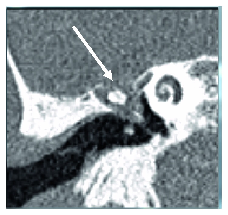
Findings: Both computerised tomography and magnetic resonance imaging of the temporal bones demonstrated a defect in the right tegmen tympani, through which a cyst herniated into the epitympanum. Postinfective changes were also noted in the right inferior temporal lobe.
Discussion: Tegmen tympani defects are a rare but important risk factor for the spread of intracranial infections from the middle ear. In cases of recurrent otogenic intracranial sepsis, it is crucial to look for evidence of this finding on imaging.
Figures

References
Publication types
LinkOut - more resources
Full Text Sources
