. 2019 Jun 26;5(6):1017-1023.
doi: 10.1021/acscentsci.9b00184. Epub 2019 May 2.

Spontaneous Formation of CdSe Photoluminescent Nanotubes with Visible-Light Photocatalytic Performance

Affiliations

Spontaneous Formation of CdSe Photoluminescent Nanotubes with Visible-Light Photocatalytic Performance

Xiaopeng Huang et al. ACS Cent Sci. .

Abstract

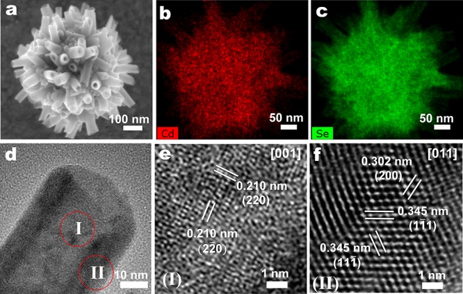
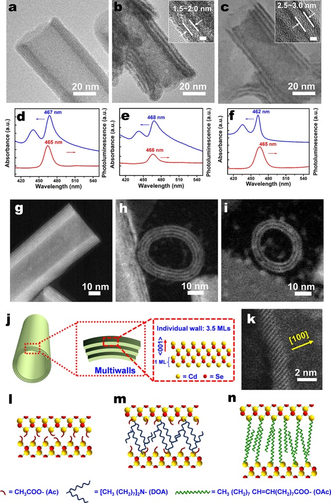
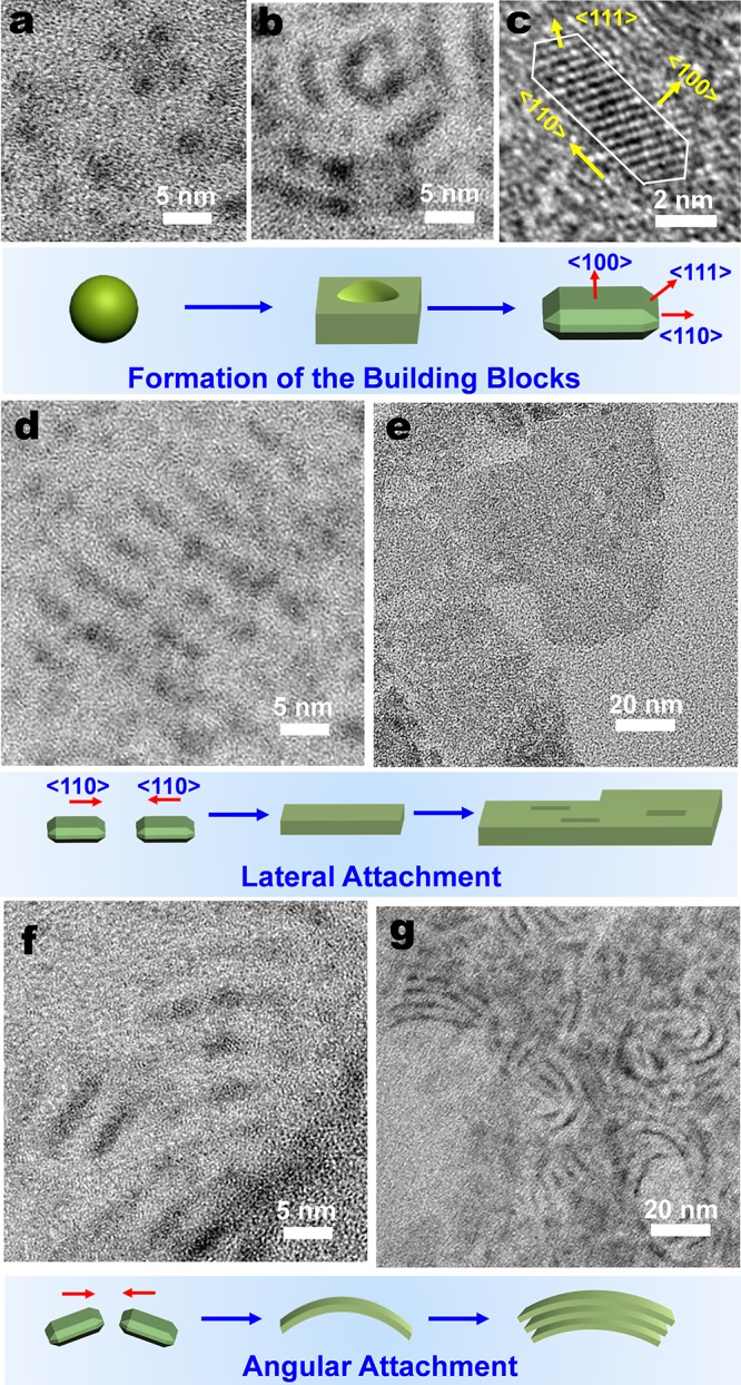
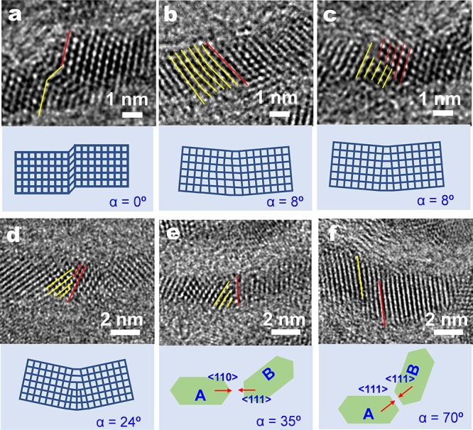
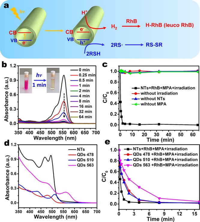
Two-dimensional (2D) colloidal CdSe nanocrystals (NCs) with precise atomic-scale thickness have attracted intensive attention in recent years due to their optical properties and quantum confinement effects originating from their particular band structure. Here, we report a solution-based and template-free protocol to synthesize CdSe nanotubes (NTs) having 3-6 walls, each of which has 3.5 molecular monolayers. Their crystal structure is zincblende, with Cd-terminated {100} planes at the top and bottom surfaces of each wall, which are passivated by short-chain acetate ligands. After verifying the prominent role of the acetate ligand for NT synthesis, we elucidated the formation mechanism of these NTs. It starts by heterogeneous nucleation of 2D plateletlike nanoseeds from the amorphous Cd precursor matrix, followed by the growth via lateral and angular attachment of nanoplatelet building blocks into curved nanosheets, eventually resulting in NTs with sharp absorption and photoluminescence peak at around 460 nm. Moreover, the NTs show remarkable visible-light photocatalytic activity, as demonstrated by the reduction of the reddish Rhodamine B into its leuco form with a conversion rate of 92% in 1 min.


Conflict of interest statement

The authors declare no competing financial interest.

Figures

Figure 1
Figure 1
Structural and elemental characterization of CdSe nanotubes (NTs) by scanning electron microscopy (SEM) and high-resolution transmission electron microscopy (HRTEM). (a) SEM graph of a bundle of CdSe NTs, with elemental mapping of (b) Cd and (c) Se. (d) HRTEM graph of a CdSe NT, with (e, f) close-ups of the center (e, part I in d), showing 0.210 nm lattice spacing corresponding to the {220} planes of the CdSe zincblende structure, and the edge (f, part II in d), showing 0.302 and 0.345 nm lattice spacings corresponding to the {200} and {111} planes, respectively, indicating that the growth direction along the thickness of the NT is ⟨100⟩.
Figure 2
Figure 2
Multiwalled character of the CdSe NTs, as observed by TEM, STEM, PL, and UV–vis absorption spectroscopy. TEM micrographs of CdSe NTs, synthesized from Cd acetate-dioctylamine (Cd(Ac)2-DOA) and Se-octadecene (Se-ODE) precursor solutions at 260 °C during a reaction time of (a) 8 min, (b) 256 min, and (c) 8 min, with the additional exchange of Ac ligands by oleic acid (OAc) at 200 °C in part c. (d–f) UV–vis absorbance and PL spectra of a solution in hexane of CdSe NTs corresponding to the structures of parts a–c, respectively. The major absorbance and PL peaks are indicative of a quasi-2D CdSe nanostructure with an individual wall thickness of 3.5 MLs. HAADF-STEM graphs of a CdSe NTs synthesized within 8 min at 260 °C when observed along the (g) tube-side direction and (h, i) tube-opening direction. (j) Schematic illustration of a multiwalled CdSe NT. (k) HAADF-STEM graph of an individual wall consisting of 3.5 MLs with ⟨100⟩ normal direction. (l–n) Schematic illustrations of the CdSe NTs corresponding to the structures of parts a–c, showing how the use of different ligands leads to different spacings between individual walls.
Figure 3
Figure 3
TEM and HRTEM study of early formation stages of CdSe NTs. (a–d, f) Synthesis at a temperature of 140 °C leads to (a) CdSe dots of around 2 nm as well as (b) seeds that form NT building blocks in the form of elongated platelets. (c) HRTEM micrograph of a small assembly of building blocks showing lattice fringe of {111} facets. The schematic shows the formation of building blocks from the CdSe dots to platelets with {100} terrace facets. (d) Lateral growth mechanism, as indicated by the attachment of the building blocks in the ⟨110⟩ direction. (e) Synthesis at a temperature of 200 °C allows the observation of holes in the nanosheets, indicative of a growth mechanism by building block assembly. (f) Angular growth mechanism by the assembly of building blocks with a rotation with respect to the ⟨110⟩ direction. (g) Formation of initial multiwalled structures as result of the angular growth mechanism and intermolecular forces between individual walls.
Figure 4
Figure 4
HRTEM study of the angular assembly mechanism. (a) HRTEM image of a distorted lattice plane due to a stacking fault along the ⟨111⟩ direction, not resulting in a microscopic rotation of the crystallographic orientation (α = 0°). (b) A small angular rotation (α = 8°) is caused by a lattice distortion on the {111} facets. Edge dislocations involving (c) one or (d) three half atomic planes into the {111} facts are also observed, contributing to an angular rotation of 8° and 24°, respectively. The oriented attachment between (e) {110} and {111} planes as well as (f) two adjacent {111} planes leads to a larger angular rotation of 35° and 70°, respectively. The red and yellow lines indicate the {111} facets in adjacent building blocks.
Figure 5
Figure 5
Visible-light photocatalytic reduction of RhB into its leuco form by using the CdSe NTs as photocatalysts. (a) Schematic illustration of the photoreduction of aqueous Rhodamine B (RhB). Under visible-light irradiation, CdSe NTs have holes in the valence band (VB) and electrons in the conduction band (CB). The former can oxidize thiols into thiolate radicals, which finally turn into more stable disulfides; the latter can reduce the protons into hydrogen, which further react with RhB to produce its leuco form. (b) Photoreduction of the RhB in the presence of CdSe NTs and 3-mercaptopropionic acid (MPA) at different light irradiation time. The inset shows the rapid decolorization of RhB after irradiation for 1 min. (c) Temporal evolution of the concentration of RhB in the presence of the NTs, MPA, and light irradiation. Control experiments in the absence of NTs, MPA, or light irradiation are also shown. (d) Absorption spectra of as-synthesized CdSe NTs and three quantum dots (QDs 478, QDs 510, and QDs 563) samples in water. The concentrations are all 0.104 mg/mL. (e) Temporal evolution of the RhB concentration during the photoreduction using CdSe NTs, QDs 478, QDs 533, and QDs 563 as the photocatalysts. The concentrations of the catalysts are all 0.05 mg/mL.

References

    1. Peng X.; Manna L.; Yang W.; Wickham J.; Scher E.; Kadavanich A.; Alivisatos A. P. Shape control of CdSe nanocrystals. Nature 2000, 404 (6773), 59.10.1038/35003535. - DOI - PubMed
    1. Manna L.; Milliron D. J.; Meisel A.; Scher E. C.; Alivisatos A. P. Controlled growth of tetrapod-branched inorganic nanocrystals. Nat. Mater. 2003, 2 (6), 382–385. 10.1038/nmat902. - DOI - PubMed
    1. Joo J.; Son J. S.; Kwon S. G.; Yu J. H.; Hyeon T. Low-temperature solution-phase synthesis of quantum well structured CdSe nanoribbons. J. Am. Chem. Soc. 2006, 128 (17), 5632–5633. 10.1021/ja0601686. - DOI - PubMed
    1. Ithurria S.; Dubertret B. Quasi 2D colloidal CdSe platelets with thicknesses controlled at the atomic level. J. Am. Chem. Soc. 2008, 130 (49), 16504–16505. 10.1021/ja807724e. - DOI - PubMed
    1. Ithurria S.; Tessier M. D.; Mahler B.; Lobo R. P.; Dubertret B.; Efros A. L. Colloidal nanoplatelets with two-dimensional electronic structure. Nat. Mater. 2011, 10 (12), 936–941. 10.1038/nmat3145. - DOI - PubMed