

Microstructural characterization of corticospinal tract in subacute and chronic stroke patients with distal lesions by means of advanced diffusion MRI
- PMID: 31263922
- PMCID: PMC6689031
- DOI: 10.1007/s00234-019-02249-2
Microstructural characterization of corticospinal tract in subacute and chronic stroke patients with distal lesions by means of advanced diffusion MRI
Abstract
Purpose: The aim of the paper is to evaluate if advanced dMRI techniques, including diffusion kurtosis imaging (DKI) and neurite orientation dispersion and density imaging (NODDI), could provide novel insights into the subtle microarchitectural modifications occurring in the corticospinal tract (CST) of stroke patients in subacute and chronic phases.
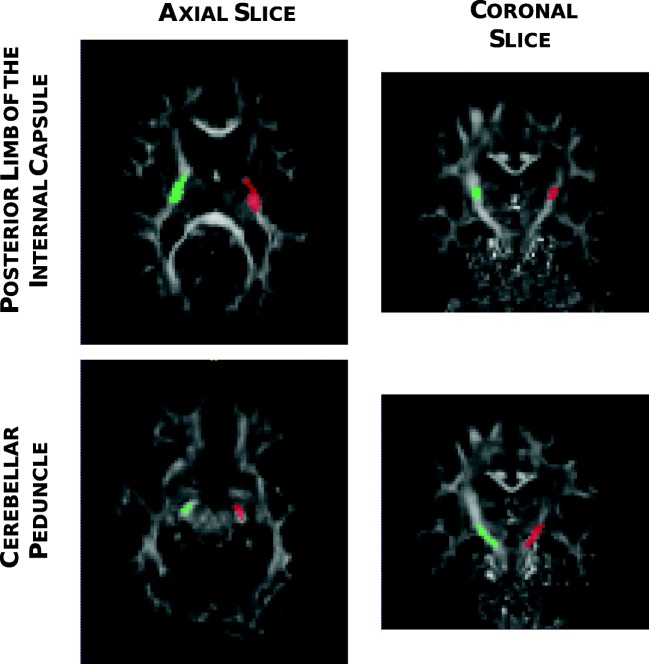
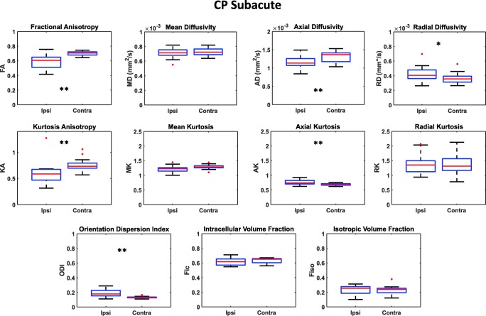
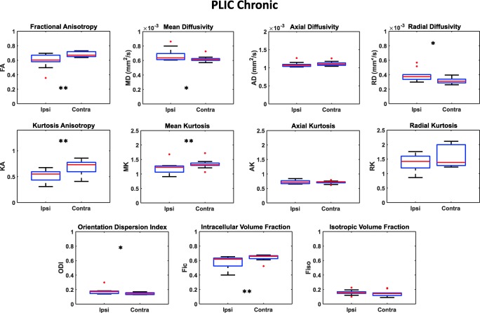
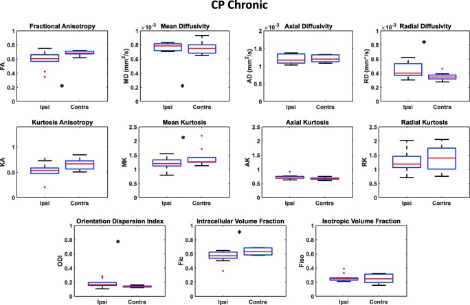
Methods: Seventeen subjects (age 68 ± 11 years) in the subacute phase (14 ± 3 days post-stroke), 10 of whom rescanned in the chronic phase (231 ± 36 days post-stroke), were enrolled. Images were acquired using a 3-T MRI scanner with a two-shell EPI protocol (20 gradient directions, b = 700 s/mm2, 3 b = 0; 64 gradient directions, b = 2000 s/mm2, 9 b = 0). DTI-, DKI-, and NODDI-derived parameters were calculated in the posterior limb of the internal capsule (PLIC) and in the cerebral peduncle (CP).
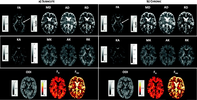
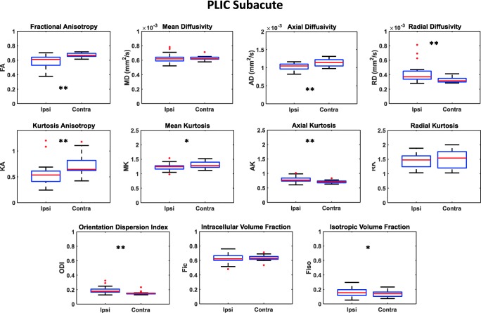
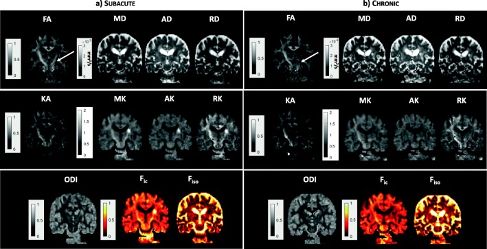
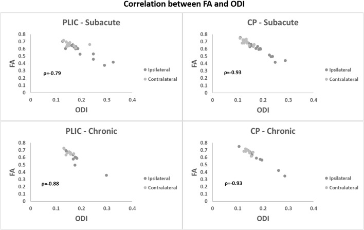
Results: In the subacute phase, a reduction of FA, AD, and KA values was correlated with an increase of ODI, RD, and AK parameters, in both the ipsilesional PLIC and CP, suggesting that increased fiber dispersion can be the main structural factor. In the chronic phase, a reduction of FA and an increase of ODI persisted in the ipsilesional areas. This was associated with reduced Fic and increased MD, with a concomitant reduction of MK and increase of RD, suggesting that fiber reduction, possibly due to nerve degeneration, could play an important role.
Conclusions: This study shows that advanced dMRI approaches can help elucidate the underpinning architectural modifications occurring in the CST after stroke. Further follow-up studies on bigger cohorts are needed to evaluate if DKI- and NODDI-derived parameters might be proposed as complementary biomarkers of brain microstructural alterations.
Keywords: Corticospinal tract; DKI; DTI; NODDI; Subacute and chronic stroke.
Conflict of interest statement
The authors declare that they have no conflict of interest.
Figures
References
-
- Hay SI, Abajobir AA, Abate KH, et al. Global, regional, and national disability-adjusted life-years (DALYs) for 333 diseases and injuries and healthy life expectancy (HALE) for 195 countries and territories, 1990–2016: a systematic analysis for the Global Burden of Disease Study 2016. Lancet. 2017;390:1260–1344. doi: 10.1016/S0140-6736(17)32130-X. - DOI - PMC - PubMed
-
- Schellinger PD, Bryan RN, Caplan LR, et al (2010) Evidence-based guideline: the role of diffusion and perfusion MRI for the diagnosis of acute ischemic stroke: report of the Therapeutics and Technology Assessment Subcommittee of the American Academy of Neurology. Neurology 75:177–185. 10.1212/WNL.0b013e3181e7c9dd - PMC - PubMed
MeSH terms
Grants and funding
- KMN142, within the project "Rete IRCCS di Neuroscienze e Neuroriabilitazione Progetto imaging/Ministero della Salute
- KMN153 within the project "Rete di Neuroimaging fase II: Ottimizzazione e armonizzazione di sequenze RM avanzate e loro applicazione nello studio delle demenze e della disabilità intellettiva in età pediatrica"/Ministero della Salute
- 5x1000 (2016 - 2017) within the project "Sviluppo di indici prognostici di imaging neuroradiologico avanzato e bio-umorali in pazienti con ictus ischemico o emorragico in fase subacuta"/IRCCS Humanitas
LinkOut - more resources
Full Text Sources
Other Literature Sources
Medical
Miscellaneous
