Muscle spindle reinnervation using transplanted embryonic dorsal root ganglion cells after peripheral nerve transection in rats
- PMID: 31264327
- PMCID: PMC6797520
- DOI: 10.1111/cpr.12660
Muscle spindle reinnervation using transplanted embryonic dorsal root ganglion cells after peripheral nerve transection in rats
Abstract
Objectives: Muscle spindles are proprioceptive receptors in the skeletal muscle. Peripheral nerve injury results in a decreased number of muscle spindles and their morphologic deterioration. However, the muscle spindles recover when skeletal muscles are reinnervated with surgical procedures, such as nerve suture or nerve transfer. Morphological changes in muscle spindles by cell transplantation procedure have not been reported so far. Therefore, we hypothesized that transplantation of embryonic sensory neurons may improve sensory neurons in the skeletal muscle and reinnervate the muscle spindles.
Materials and methods: We collected sensory neurons from dorsal root ganglions of 14-day-old rat embryos and prepared a rat model of peripheral nerve injury by performing sciatic nerve transection and allowing for a period of one week before which we performed the cell transplantations. Six months later, the morphological changes of muscle spindles in the cell transplantation group were compared with the naïve control and surgical control groups.
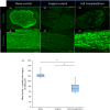
Results: Our results demonstrated that transplantation of embryonic dorsal root ganglion cells induced regeneration of sensory nerve fibre and reinnervation of muscle spindles in the skeletal muscle. Moreover, calbindin D-28k immunoreactivity in intrafusal muscle fibres was maintained for six months after denervation in the cell transplantation group, whereas it disappeared in the surgical control group.
Conclusions: Cell transplantation therapies could serve as selective targets to modulate mechanosensory function in the skeletal muscle.
© 2019 The Authors. Cell Proliferation Published by John Wiley & Sons Ltd.
Conflict of interest statement
The authors have declared that there is no conflict of interest.
Figures


Similar articles
-
Innervation of Meissner's corpuscles and Merkel -cells by transplantation of embryonic dorsal root ganglion cells after peripheral nerve section in rats.J Tissue Eng Regen Med. 2021 Jun;15(6):586-595. doi: 10.1002/term.3196. Epub 2021 Apr 23. J Tissue Eng Regen Med. 2021. PMID: 33837671
-
Calbindin D-28k-immunoreactivity in rat muscle spindles during postnatal maturation and after denervation.Histochem J. 1992 Sep;24(9):673-8. doi: 10.1007/BF01047588. Histochem J. 1992. PMID: 1429002
-
Sensory protection of rat muscle spindles following peripheral nerve injury and reinnervation.Plast Reconstr Surg. 2009 Dec;124(6):1860-1868. doi: 10.1097/PRS.0b013e3181bcee47. Plast Reconstr Surg. 2009. PMID: 19952642 Free PMC article.
-
Influence of peripheral and central targets on subpopulations of sensory neurons expressing calbindin immunoreactivity in the dorsal root ganglion of the chick embryo.Neuroscience. 1988 Jul;26(1):225-32. doi: 10.1016/0306-4522(88)90139-x. Neuroscience. 1988. PMID: 3419587
-
Sensory nerve regeneration and reinnervation in muscle following peripheral nerve injury.Muscle Nerve. 2022 Oct;66(4):384-396. doi: 10.1002/mus.27661. Epub 2022 Jul 2. Muscle Nerve. 2022. PMID: 35779064 Review.
Cited by
-
(-)-Epigallocatechin gallate-loaded polycaprolactone scaffolds fabricated using a 3D integrated moulding method alleviate immune stress and induce neurogenesis.Cell Prolif. 2020 Jan;53(1):e12730. doi: 10.1111/cpr.12730. Epub 2019 Nov 20. Cell Prolif. 2020. PMID: 31746040 Free PMC article.
-
Distribution Heterogeneity of Muscle Spindles Across Skeletal Muscles of Lower Extremities in C57BL/6 Mice.Front Neuroanat. 2022 Mar 17;16:838951. doi: 10.3389/fnana.2022.838951. eCollection 2022. Front Neuroanat. 2022. PMID: 35370570 Free PMC article.
-
Evaluating Sexual Dimorphism of the Muscle Spindles and Intrafusal Muscle Fibers in the Medial Gastrocnemius of Male and Female Rats.Front Neuroanat. 2021 Oct 1;15:734555. doi: 10.3389/fnana.2021.734555. eCollection 2021. Front Neuroanat. 2021. PMID: 34658799 Free PMC article.
-
Functional Reconstruction of Denervated Muscle by Xenotransplantation of Neural Cells from Porcine to Rat.Int J Mol Sci. 2022 Aug 7;23(15):8773. doi: 10.3390/ijms23158773. Int J Mol Sci. 2022. PMID: 35955906 Free PMC article.
References
-
- Bain JR, Veltri KL, Chamberlain D, Fahnestock M. Improved functional recovery of denervated skeletal muscle after temporary sensory nerve innervation. Neuroscience. 2001;103:503‐510. - PubMed
-
- Mosahebi A, Fuller P, Wiberg M, Terenghi G. Effect of allogeneic Schwann cell transplantation on peripheral nerve regeneration. Exp Neurol. 2002;173:213‐223. - PubMed
MeSH terms
Substances
LinkOut - more resources
Full Text Sources
Medical

