

Antimicrobial effects of microwave-induced plasma torch (MiniMIP) treatment on Candida albicans biofilms
- PMID: 31264377
- PMCID: PMC6680639
- DOI: 10.1111/1751-7915.13459
Antimicrobial effects of microwave-induced plasma torch (MiniMIP) treatment on Candida albicans biofilms
Abstract
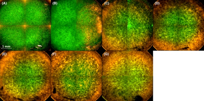
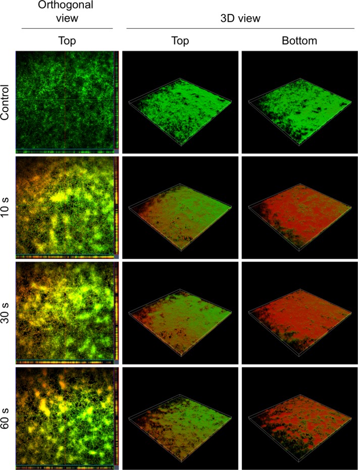
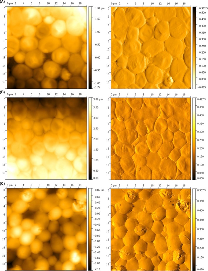
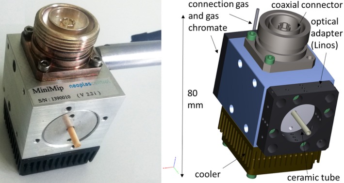
The susceptibility of Candida albicans biofilms to a non-thermal plasma treatment has been investigated in terms of growth, survival and cell viability by a series of in vitro experiments. For different time periods, the C. albicans strain SC5314 was treated with a microwave-induced plasma torch (MiniMIP). The MiniMIP treatment had a strong effect (reduction factor (RF) = 2.97 after 50 s treatment) at a distance of 3 cm between the nozzle and the superior regions of the biofilms. In addition, a viability reduction of 77% after a 20 s plasma treatment and a metabolism reduction of 90% after a 40 s plasma treatment time were observed for C. albicans. After such a treatment, the biofilms revealed an altered morphology of their cells by atomic force microscopy (AFM). Additionally, fluorescence microscopy and confocal laser scanning microscopy (CLSM) analyses of plasma-treated biofilms showed that an inactivation of cells mainly appeared on the bottom side of the biofilms. Thus, the plasma inactivation of the overgrown surface reveals a new possibility to combat biofilms.
© 2019 The Authors. Microbial Biotechnology published by John Wiley & Sons Ltd and Society for Applied Microbiology.
Conflict of interest statement
None declared.
Figures


Similar articles
-
Nonthermal Plasma Jet Treatment Negatively Affects the Viability and Structure of Candida albicans SC5314 Biofilms.Appl Environ Microbiol. 2018 Oct 17;84(21):e01163-18. doi: 10.1128/AEM.01163-18. Print 2018 Nov 1. Appl Environ Microbiol. 2018. PMID: 30143511 Free PMC article.
-
In vitro treatment of Candida albicans biofilms on denture base material with volume dielectric barrier discharge plasma (VDBD) compared with common chemical antiseptics.Clin Oral Investig. 2015 Dec;19(9):2319-26. doi: 10.1007/s00784-015-1463-y. Epub 2015 Apr 22. Clin Oral Investig. 2015. PMID: 25898894
-
Contact-free inactivation of Candida albicans biofilms by cold atmospheric air plasma.Appl Environ Microbiol. 2012 Jun;78(12):4242-7. doi: 10.1128/AEM.07235-11. Epub 2012 Mar 30. Appl Environ Microbiol. 2012. PMID: 22467505 Free PMC article.
-
Microscopical analysis of Candida albicans biofilms on heat-polymerised acrylic resin after chlorhexidine gluconate and sodium hypochlorite treatments.Mycoses. 2011 Nov;54(6):e712-7. doi: 10.1111/j.1439-0507.2010.02005.x. Epub 2011 May 23. Mycoses. 2011. PMID: 21605193
-
Effect of curcumin-mediated photodynamic therapy on Streptococcus mutans and Candida albicans: A systematic review of in vitro studies.Photodiagnosis Photodyn Ther. 2019 Sep;27:455-461. doi: 10.1016/j.pdpdt.2019.07.010. Epub 2019 Jul 25. Photodiagnosis Photodyn Ther. 2019. PMID: 31352059
Cited by
-
Evaluation of the biofilm life cycle between Candida albicans and Candida tropicalis.Front Cell Infect Microbiol. 2022 Aug 18;12:953168. doi: 10.3389/fcimb.2022.953168. eCollection 2022. Front Cell Infect Microbiol. 2022. PMID: 36061861 Free PMC article.
-
Inhibition of polymicrobial biofilm formation by saw palmetto oil, lauric acid and myristic acid.Microb Biotechnol. 2022 Feb;15(2):590-602. doi: 10.1111/1751-7915.13864. Epub 2021 Jun 22. Microb Biotechnol. 2022. PMID: 34156757 Free PMC article.
-
Physics Comes to the Aid of Medicine-Clinically-Relevant Microorganisms through the Eyes of Atomic Force Microscope.Pathogens. 2020 Nov 20;9(11):969. doi: 10.3390/pathogens9110969. Pathogens. 2020. PMID: 33233696 Free PMC article. Review.
-
Plasma-Treated Water: A Comparison with Analog Mixtures of Traceable Ingredients.Microorganisms. 2023 Apr 3;11(4):932. doi: 10.3390/microorganisms11040932. Microorganisms. 2023. PMID: 37110355 Free PMC article.
-
Appraisal of Cinnamaldehyde Analogs as Dual-Acting Antibiofilm and Anthelmintic Agents.Front Microbiol. 2022 Mar 16;13:818165. doi: 10.3389/fmicb.2022.818165. eCollection 2022. Front Microbiol. 2022. PMID: 35369516 Free PMC article.
References
-
- Al‐Fattani, M.A. , and Douglas, L.J. (2006) Biofilm matrix of Candida albicans and Candida tropicalis: chemical composition and role in drug resistance. J Med Microbiol 55: 999–1008. - PubMed
-
- Anderson, J.B. (2005) Evolution of antifungal‐drug resistance: mechanisms and pathogen fitness. Nat Rev Microbiol 3: 547–556. - PubMed
-
- Baeva, M. , Bösel, A. , Ehlbeck, J. , and Loffhagen, D. (2012) Modeling of microwave‐induced plasma in argon at atmospheric pressure. Phys Rev E 85: 056404. - PubMed
-
- Baillie, G.S. , and Douglas, L.J. (2000) Matrix polymers of Candida biofilms and their possible role in biofilm resistance to antifungal agents. J Antimicrob Chemother 46: 397–403. - PubMed
Publication types
MeSH terms
Substances
LinkOut - more resources
Full Text Sources
Miscellaneous
