. 2020 Jan 10;30(1):382-390.
doi: 10.1093/cercor/bhz094.

Quantitative MRI Analyses of Regional Brain Growth in Living Fetuses with Down Syndrome

Affiliations

Quantitative MRI Analyses of Regional Brain Growth in Living Fetuses with Down Syndrome

Tomo Tarui et al. Cereb Cortex. .

Abstract

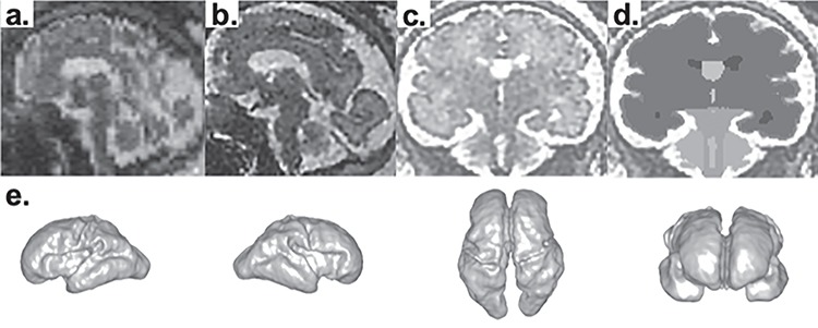
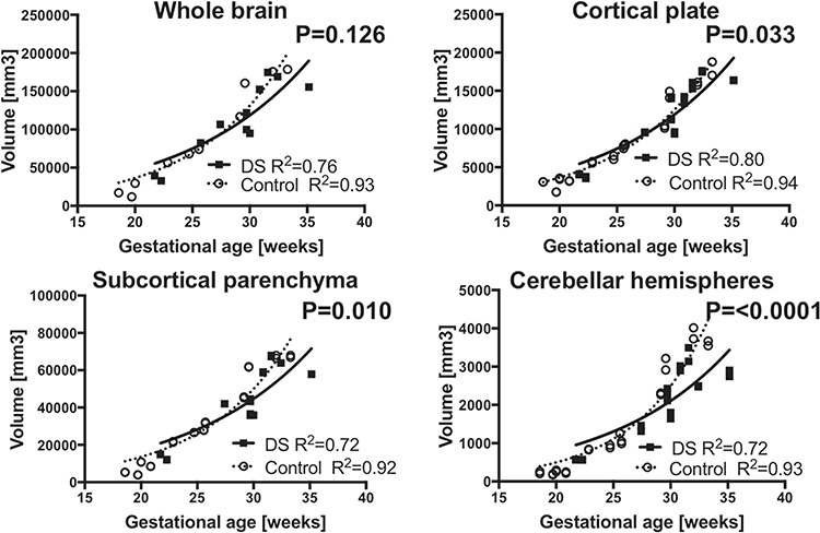
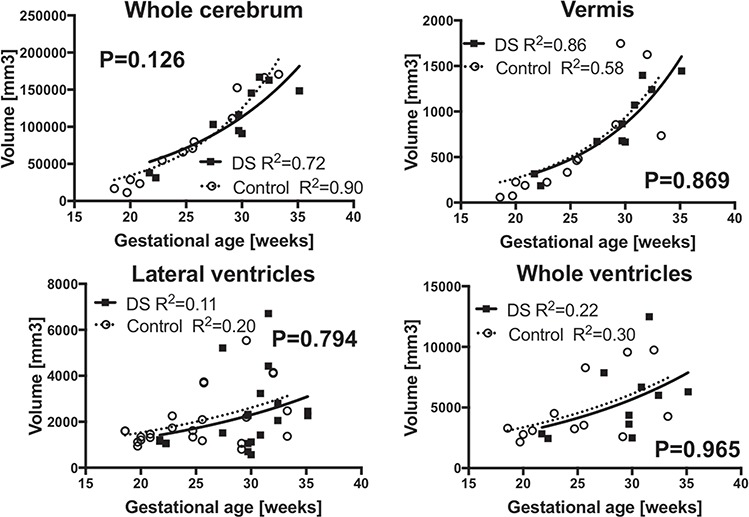
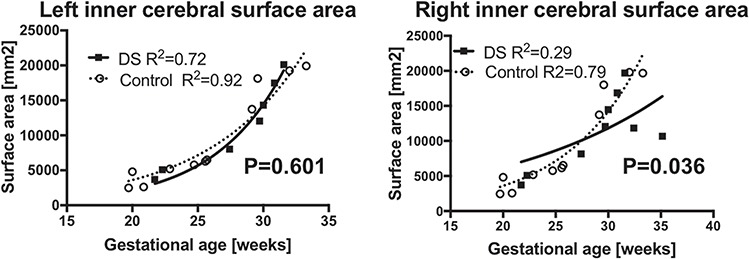
Down syndrome (DS) is the most common liveborn autosomal chromosomal anomaly and is a major cause of developmental disability. Atypical brain development and the resulting intellectual disability originate during the fetal period. Perinatal interventions to correct such aberrant development are on the horizon in preclinical studies. However, we lack tools to sensitively measure aberrant structural brain development in living human fetuses with DS. In this study, we aimed to develop safe and precise neuroimaging measures to monitor fetal brain development in DS. We measured growth patterns of regional brain structures in 10 fetal brains with DS (29.1 ± 4.2, weeks of gestation, mean ± SD, range 21.7~35.1) and 12 control fetuses (25.2 ± 5.0, range 18.6~33.3) using regional volumetric analysis of fetal brain MRI. All cases with DS had confirmed karyotypes. We performed non-linear regression models to compare fitted regional growth curves between DS and controls. We found decreased growth trajectories of the cortical plate (P = 0.033), the subcortical parenchyma (P = 0.010), and the cerebellar hemispheres (P < 0.0001) in DS compared to controls. This study provides proof of principle that regional volumetric analysis of fetal brain MRI facilitates successful evaluation of brain development in living fetuses with DS.

Keywords: Down syndrome; brain development; fetal MRI; human fetus; volumetric analysis.


Figures

Figure 1
Figure 1
Processing fetal MRI for surface analysis. Raw volume data of fetal MRI (a) is processed with motion correction and high-resolution volume reconstruction to reconstruct 3D fetal brain image (b). In coronal images of reconstructed volume images (c), the regional structures of the brain—cortical plate, subcortical parenchyma, cerebellar hemispheres, vermis, brainstem, lateral-, third-, and fourth ventricles—were manually segmented on each slice using Freeview (d). 3D volume rendering of segmentation data of MRI DS08b (e).
Figure 2
Figure 2
Distinct regional growth trajectories in fetuses with DS and CT. Growth trajectories of regional volume were modeled according to non-linear regression models and compared between fetuses with DS and CT. Growth trajectory of cortical plate (b), subcortical parenchyma (c), and cerebellar hemispheres (d) were significantly smaller in fetuses with DS compared to the CT. Measures from left and right cortical plate, subcortical parenchyma, and cerebellar hemispheres were separately plotted. Growth trajectory of whole brain (a) is in different between DS and CT. Volumes of cortical plate, subcortical parenchyma, and cerebellar hemispheres from each hemisphere were plotted separately.
Figure 3
Figure 3
Similar regional growth trajectories in fetuses with DS and CT. Growth trajectory of whole cerebrum (a), vermis (b), lateral ventricles (c), and whole ventricles (d) were indifferent between fetuses with DS and the CT. Measures from left and right lateral ventricles were separately plotted.
Figure 4
Figure 4
Inner cerebral surface area. Growth trajectory of inner cerebral surface was smaller in right hemispheres (b) in fetuses with DS. However, inner surface area of the left hemispheres (a) were indifferent between DS and the CT.

References

    1. Aylward EH, Li Q, Honeycutt NA, Warren AC, Pulsifer MB, Barta PE, Chan MD, Smith PD, Jerram M, Pearlson GD. 1999. MRI volumes of the hippocampus and amygdala in adults with Down's syndrome with and without dementia. Am J Psychiatry. 156:564–568. - PubMed
    1. Benkarim OM, Hahner N, Piella G, Gratacos E, Gonzalez Ballester MA, Eixarch E, Sanroma G. 2018. Cortical folding alterations in fetuses with isolated non-severe ventriculomegaly. Neuroimage Clin. 18:103–114. - PMC - PubMed
    1. Bhattacharyya A, McMillan E, Chen SI, Wallace K, Svendsen CN. 2009. A critical period in cortical interneuron neurogenesis in down syndrome revealed by human neural progenitor cells. Dev Neurosci. 31:497–510. - PMC - PubMed
    1. Bianchi DW, Chiu RWK. 2018. Sequencing of circulating cell-free DNA during pregnancy. N Engl J Med. 379:464–473. - PMC - PubMed
    1. Biegon A, Hoffmann C. 2014. Quantitative magnetic resonance imaging of the fetal brain in utero: methods and applications. World J Radiol. 6:523–529. - PMC - PubMed

Publication types