. 2019 Aug;40(8):1115-1126.
doi: 10.1002/humu.23760. Epub 2019 May 5.

Heterozygous variants in MYBPC1 are associated with an expanded neuromuscular phenotype beyond arthrogryposis

Collaborators, Affiliations

Heterozygous variants in MYBPC1 are associated with an expanded neuromuscular phenotype beyond arthrogryposis

Vandana Shashi et al. Hum Mutat. 2019 Aug.

Abstract

Encoding the slow skeletal muscle isoform of myosin binding protein-C, MYBPC1 is associated with autosomal dominant and recessive forms of arthrogryposis. The authors describe a novel association for MYBPC1 in four patients from three independent families with skeletal muscle weakness, myogenic tremors, and hypotonia with gradual clinical improvement. The patients carried one of two de novo heterozygous variants in MYBPC1, with the p.Leu263Arg variant seen in three individuals and the p.Leu259Pro variant in one individual. Both variants are absent from controls, well conserved across vertebrate species, predicted to be damaging, and located in the M-motif. Protein modeling studies suggested that the p.Leu263Arg variant affects the stability of the M-motif, whereas the p.Leu259Pro variant alters its structure. In vitro biochemical and kinetic studies demonstrated that the p.Leu263Arg variant results in decreased binding of the M-motif to myosin, which likely impairs the formation of actomyosin cross-bridges during muscle contraction. Collectively, our data substantiate that damaging variants in MYBPC1 are associated with a new form of an early-onset myopathy with tremor, which is a defining and consistent characteristic in all affected individuals, with no contractures. Recognition of this expanded myopathic phenotype can enable identification of individuals with MYBPC1 variants without arthrogryposis.

Keywords: MYBPC1; arthrogryposis; hypotonia; myopathy; myosin binding protein-C; tremor.


Conflict of interest statement

CONFLICT OF INTEREST STATEMENT

None of the authors has a conflict of interest.

Figures

Figure 1:
Figure 1:
Pathogenic variants in MYBPC1. (A) Schematic representation of the domain structure of sMyBP-C encoded by MYBPC1 and depiction of known (in blue) and novel (in red) pathogenic variants; Ig and FN-III domains are shown as white and grey rectangles, the Pro/Ala rich motif and the M-motif are illustrated as dark and light grey, thick, horizontal lines, and spliced sequences are depicted as colored, perpendicular lines. (B) Pedigrees and Sanger traces of the three families containing the novel pathogenic MYBPC1 variants. Individuals 1, 3 and 4 carry the p.Leu263Arg variant, which appeared de novo in individuals 1 and 4 belonging in two different families, and was inherited by individual 3 (daughter; proband) from individual 4 (father), while individual 2 belonging to a third unrelated family carries the de novo p.Leu259Pro variant. (C) Evolutionary conservation of L259 and L263 across orthologs from vertebrate species; conserved residues surrounding L259 and L263 within the M-motif are highlighted in yellow, while L259 and L263 are marked with red boxes; H.s. denotes Homo sapiens; M.m., Mus musculus; O.c., Oryctolagus cuniculus; B.t., Bos taurus; G.g., Gallus gallus; X.t., Xenopus tropicalis; D.r., Danio rerio.
Figure 2:
Figure 2:
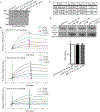
(A) Coomassie blue stained gel showing equivalent amounts (1.5 μg) of control GST, wild-type GST-P/A-C1-M, mutant GST-P/A-C1-M L259P, and mutant GST-P/A-C1-M L263R proteins used in the overlay and Biacore assays. Full length GST-P/A-C1-M recombinant proteins (~58 kDa) are denoted with an arrow; the preparations of both mutant proteins contain a very closely migrating degradation product (56–57 kDa), while a degradation product of ~50 kDa is present in all three protein preparations; non-continuous lanes are separated by a white line. (B) Real time kinetics evaluation of the interaction between wild-type or mutant GST-P/A-C1-M proteins and HMM using a Biacore 3000 SPR biosensor. (C) Kinetic measurements at equilibrium indicated that GST-P/A-C1-M L263R binding to HMM was decreased by ~5.5-fold compared to wild-type levels. (D) Equivalent amounts (3 μg) of purified actin were separated by SDS-PAGE, transferred to nitrocellulose membrane, and overlaid with 0.5 μg/mL of control GST-protein, wild-type GST-P/A-C1-M, or mutant GST-P/A-C1-M carrying the L259P or L263R mutation. Nitrocellulose membranes (Ponceau stain) and films (Western blots probed with GST-ab) were cropped to only include the area of expected signal. Densitometric quantification of at least three independent overlay/immunoblotting experiments indicated that neither mutation significantly affected binding to actin. Control GST-proteins did not bind actin.
Figure 3:
Figure 3:
Circular dichroism (CD) experiments and computation modeling analysis of the L259P and L263R mutations. (A) Recombinant wild-type and mutant His-tagged proteins containing the M-motif that carries the L259P and L263R mutations were used in CD experiments. CD spectra at 30 °C showed that mutant M-motif containing the L259P mutation (hollow diamonds) is ~10% less helical compared to wild-type (solid circles). (B) Representative snapshot following molecular dynamics (MD) indicated that wild-type M-motif protein (top; magenta) has normal hydrogen bonds (black dashed lines) at the beginning of the M-domain helix 3 (L259-L263; R260-K264; G261-R265). On the contrary, MD of mutant M-motif protein containing the L259P variant (bottom; cyan) showed that these hydrogen bonds are no longer well-formed (red dashed lines). (C) P259 (purple) still packs well into the hydrophobic interior (cyan spheres) of the M-motif. (D) Representative SDS-PAGE gel of trypsin digestion assays for recombinant wild-type and mutant L259P M-motif proteins stained with Coomassie Brilliant Blue, showing increased cleavage products in the presence of the L259P mutation. (E) Normalized molar ellipticity at 222 nm vs. temperature showed that mutant M-motif carrying the L263R variant (hollow diamonds) is less stable than wild-type (solid circles). (F) Representative model of wild-type (purple) and mutant M-motif containing the L263R mutation (green) after a 100 ns MD equilibration. Mutant M-motif does not pack as well into the hydrophobic interior as wild-type, but does form a previously absent favorable interaction between the R263 and E248 sidechains. This decrease in stability and alterations in intramolecular hydrogen bonds may be the cause of decreased binding to myosin seen in the in vitro binding assay.

Similar articles

Cited by

References

    1. Ackermann MA, & Kontrogianni-Konstantopoulos A (2010). Myosin binding protein-C slow: an intricate subfamily of proteins. J Biomed Biotechnol, 2010, 652065. doi:10.1155/2010/652065 - DOI - PMC - PubMed
    1. Ackermann MA, & Kontrogianni-Konstantopoulos A (2011). Myosin binding protein-C slow is a novel substrate for protein kinase A (PKA) and C (PKC) in skeletal muscle. J Proteome Res, 10(10), 4547–4555. doi:10.1021/pr200355w - DOI - PMC - PubMed
    1. Ackermann MA, & Kontrogianni-Konstantopoulos A (2013). Myosin binding protein-C slow: a multifaceted family of proteins with a complex expression profile in fast and slow twitch skeletal muscles. Front Physiol, 4, 391. doi:10.3389/fphys.2013.00391 - DOI - PMC - PubMed
    1. Adzhubei IA, Schmidt S, Peshkin L, Ramensky VE, Gerasimova A, Bork P, … Sunyaev SR (2010). A method and server for predicting damaging missense mutations. Nat Methods, 7(4), 248–249. doi:10.1038/nmeth0410-248 - DOI - PMC - PubMed
    1. Atlas, H. P. (version 17). https://www.proteinatlas.org/.

Publication types