. 2019 Sep 5;47(15):8301-8317.
doi: 10.1093/nar/gkz571.

Role of Era in assembly and homeostasis of the ribosomal small subunit

Affiliations

Role of Era in assembly and homeostasis of the ribosomal small subunit

Aida Razi et al. Nucleic Acids Res. .

Abstract

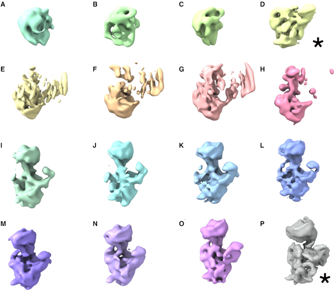
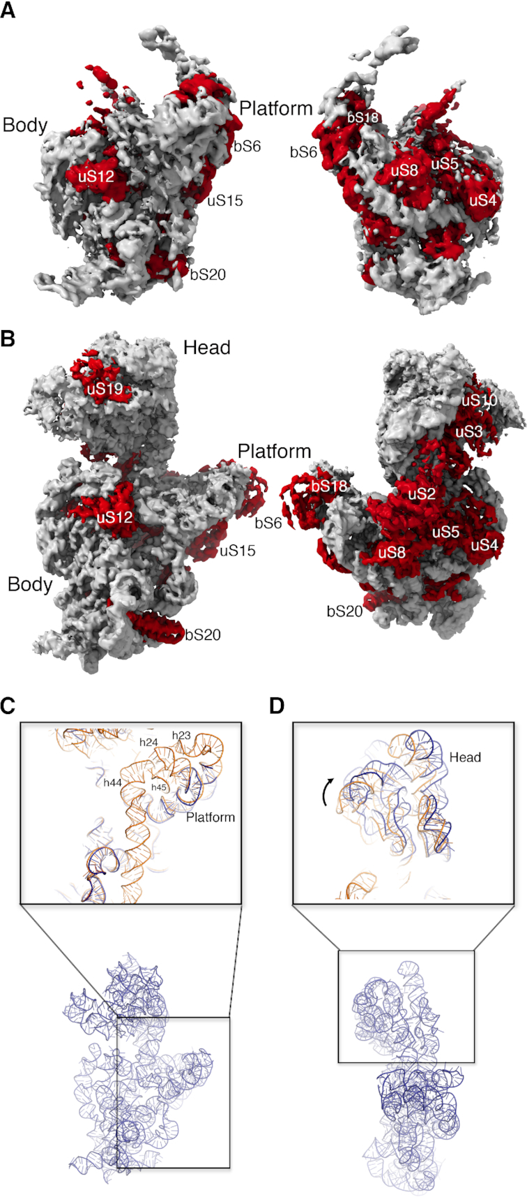
Assembly factors provide speed and directionality to the maturation process of the 30S subunit in bacteria. To gain a more precise understanding of how these proteins mediate 30S maturation, it is important to expand on studies of 30S assembly intermediates purified from bacterial strains lacking particular maturation factors. To reveal the role of the essential protein Era in the assembly of the 30S ribosomal subunit, we analyzed assembly intermediates that accumulated in Era-depleted Escherichia coli cells using quantitative mass spectrometry, high resolution cryo-electron microscopy and in-cell footprinting. Our combined approach allowed for visualization of the small subunit as it assembled and revealed that with the exception of key helices in the platform domain, all other 16S rRNA domains fold even in the absence of Era. Notably, the maturing particles did not stall while waiting for the platform domain to mature and instead re-routed their folding pathway to enable concerted maturation of other structural motifs spanning multiple rRNA domains. We also found that binding of Era to the mature 30S subunit destabilized helix 44 and the decoding center preventing binding of YjeQ, another assembly factor. This work establishes Era's role in ribosome assembly and suggests new roles in maintaining ribosome homeostasis.


Figures

Figure 1.
Figure 1.
Ribosomal proteins and assembly factors content of the 30S particles in Era depleted cells. (A) Ribosomal profiles of the Era depleted strain under Era expression (+arabinose) and Era depletion (-arabinose) conditions. The asterisk and vertical lines in the bottom gradient indicate the fractions that provided the purified 30SEra-depleted particles for the qMS and cryo-EM experiments. The pie charts indicate the fraction of ribosomal particles represented by free 30S subunits. (B) Heat map showing protein abundance of the 30S particles purified from the parental strain and the Era depleted strain grown under (-arabinose) conditions. Protein abundance is shown relative to a purified 70S particle. Occupancy patterns were hierarchically clustered revealing three groups, which are marked. Proteins in each group are labeled in a different color. Sample replicas were also hierarchically classified and clustered in two groups indicated as 30SdepEra (30SEra-depleted particles purified from Era depleted strain grown under (-arabinose) conditions) and 30SWT (30S subunits purified from parental strain). (C) Protein occupancy levels from the qMS analysis of the 30SEra-depleted particles were plotted in the Nomura assembly map of the 30S subunit using the same color coding as in panel (A). (D) The r-proteins found to be sub-stoichiometric by qMS are shown in a color different from gray in the structure of the mature 30S subunit. The entire r-protein bS1 is not shown for clarity. The oval shape labeled as bS1 indicates the binding site of the N-terminal region of this protein to the 30S subunit. Ribosomal proteins found on full occupancy in the 30SEra-depleted particles are shown in gray.
Figure 2.
Figure 2.
Immature 30S particles accumulating in the Era-depleted strain. (AP) Cryo-EM maps obtained from a sample containing purified 30SEra-depleted particles using 3D image classification approaches. The most abundant classes are marked by an asterisk. Classes D and P represented the 22% and 50% of the population, respectively. The remaining classes accumulated only a 28% of the particle population.
Figure 3.
Figure 3.
Cryo-EM structures of the most abundant 30SEra-depleted particles. (A) Front and back view of the cryo-EM map of the 30SEra-depleted particle class D. Ribosomal proteins and rRNA in the structure are colored in red and light gray, respectively. (B) Similar representation of the cryo-EM map of the 30SEra-depleted particle class P. (C) Zoom-in-view of the 3′ minor and central domains of the atomic model obtained for the 30SEra-depleted particle class P. The area framed with a black square in the overall view of the atomic model (r-proteins removed for clarity) at the bottom is shown enlarged in the top panel. The top panel shows the superimposition of the atomic model for the mature 30S subunit (orange) (PDB ID: 4V4Q) and the corresponding region on the model obtained for the 30SEra-depleted particle (dark blue). (D) Zoom-in-view of the 3′ major domain of the atomic model obtained for the 30SEra-depleted particle class P. This region of the model is compared in the top panel with the atomic model of the mature 30S subunit following the same color code as in panel (C). Ribosomal proteins have been removed for clarity.
Figure 4.
Figure 4.
In vivo DMS footprinting of Era-dependent changes in the 16S central domain. (A) Scheme of the in vivo DMS footprinting experiment after Era depletion and re-expression. (B) DMS modification patterns of different Era samples as detected by extension of primer 812 (Supplementary Table S4). The Era pre, Era depleted and Era re-expression cells were untreated (–) or modified with DMS for 0.5, 1 or 2 min. DMS modified nucleotides are highlighted with colored bars (right). The nucleotide numbers are indicated to the left of the sequencing ladder, and the unextended primer is marked (3′). (C) DMS-modified nucleotides on the 16S rRNA secondary structure are colored based on the effect of Era as indicated in the key and illustrated in Supplementary Figure S5. The primer binding location is shaded gray.
Figure 5.
Figure 5.
Era dissociates 70S ribosomes and destabilizes helix 44 and decoding center. (A) Ribosome profiles obtained during the 70S dissociation experiments. Plot lines from the reaction containing GMPPNP, GTP, GDP or no nucleotide are shown. All reactions contained a 5-fold excess of Era with respect to the 70S ribosomes. (B) Comparison of the cryo-EM map obtained for the Era-treated 30S (class II) (left panel) with the structure of the mature 30S subunit (PDB ID: 4V4Q) (right panel). The r-RNA and r-proteins are labeled and colored in grey and orange, respectively in both structures. Structural elements absent in the cryo-EM map obtained for the 30S particles treated with Era are colored in blue in the structure of the mature 30S subunit. This panel shows a front view of the structures. (C) Comparison of the back view of the cryo-EM map obtained for the Era treated 30S particles (class II) (left panel) with the structure of the mature 30S subunit (right panel) using the same coloring scheme as in panel (B). For clarity, bS1 is not shown in the mature 30S subunit. The fragmented density potentially representing Era is indicated with an arrow and colored in cyan.
Figure 6.
Figure 6.
Era blocks binding of YjeQ to the 30S subunit. (A) Analysis of the interaction of YjeQ with the mature 30S subunit by MST. Ribosomal particles were fluorescently labeled and maintained at constant concentration in the three assays. In one assay, the ribosomal subunits were present by themselves and unlabeled YjeQ was titrated into the reaction (top panels). In the other two assays, before YjeQ was added at increasing concentrations, the mature 30S subunits were first mixed with a 10-fold (middle panels) or 20-fold excess of Era (bottom panels). After a short incubation with YjeQ, reactions were loaded into the capillaries and the MST signal of the labeled ribosomal particles (left panels) was measured. Measured changes in the MST response were used to produce curves that plotted the Fnorm (%) = F1/F0 versus the logarithm of YjeQ concentration. The F1 and F0 regions of the fluorescence time traces used to calculate Fnorm (%) are indicated in the panel (A). (B) Front and (C) back views of the cryo-EM map obtained for the Era+YjeQ-treated 30S (class II) (left panels). Ribosomal proteins and rRNA in the structure are colored in green and light gray, respectively. Equivalent views of the mature 30S subunit are shown in the right panels with the structural components displayed using the same color scheme. Structural elements absent in the cryo-EM map obtained for the 30S particles treated with Era and YjeQ are colored in blue in the structure of the mature 30S subunit. For clarity, bS1 is not shown in the mature 30S subunit.
Figure 7.
Figure 7.
Era depletion causes re-routing of the assembling 30S particles. Diagram summarizing the effect of Era depletion on the assembly of 30S subunits. Cellular depletion of the essential assembly factor Era does not stall the assembly of the 30S subunit, causing accumulation of assembly intermediates upstream of the reaction catalyzed by Era. Rather, in the absence of the Era, the 30S particles are unable to fold helices 23 and 24 in the platform domain; however, they re-route their folding pathway and continue the maturation of other structural motifs. During this process, all of the 16S rRNA domains of the 30S subunit fold independently and simultaneously accumulating a variety of assembly intermediates that were characterized in this study using cryo-EM. The red asterisks indicate that helices 23 and 24 still adopt an immature conformation in all of these structures. Only re-introduction of Era in the cells allow these assembly intermediate to reach a mature state. The ribosomal particles shown as gray flat profiles indicate the physiological assembly intermediates that exist in the cell when Era is present at normal concentrations. These particles are capable of folding helices 23 and 24 normally and reach the mature state.

Similar articles

Cited by

References

    1. Adilakshmi T., Ramaswamy P., Woodson S.A.. Protein-independent folding pathway of the 16S rRNA 5′ domain. J. Mol. Biol. 2005; 351:508–519. - PubMed
    1. Talkington M.W., Siuzdak G., Williamson J.R.. An assembly landscape for the 30S ribosomal subunit. Nature. 2005; 438:628–632. - PMC - PubMed
    1. Shajani Z., Sykes M.T., Williamson J.R.. Assembly of bacterial ribosomes. Annu. Rev. Biochem. 2011; 80:501–526. - PubMed
    1. Leipe D.D., Wolf Y.I., Koonin E.V., Aravind L.. Classification and evolution of P-loop GTPases and related ATPases. J. Mol. Biol. 2002; 317:41–72. - PubMed
    1. Takiff H.E., Chen S.M., Court D.L.. Genetic analysis of the rnc operon of Escherichia coli. J. Bacteriol. 1989; 171:2581–2590. - PMC - PubMed

Publication types

MeSH terms