. 2019 Aug;15(34):e1900968.
doi: 10.1002/smll.201900968. Epub 2019 Jul 2.

Targeted Gold Nanocluster-Enhanced Radiotherapy of Prostate Cancer

Affiliations

Targeted Gold Nanocluster-Enhanced Radiotherapy of Prostate Cancer

Dong Luo et al. Small. 2019 Aug.

Abstract

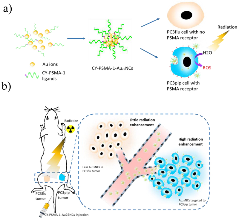
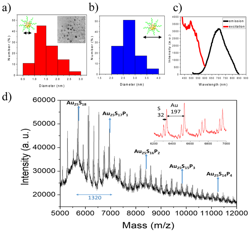
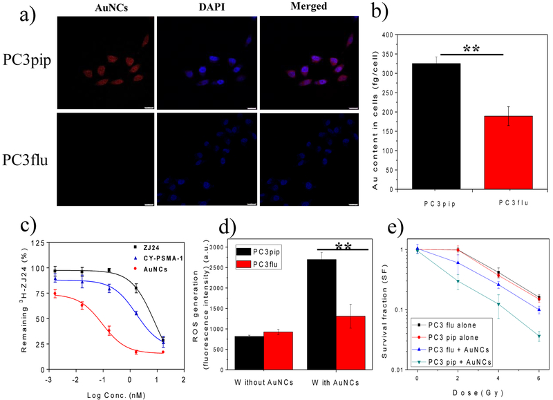
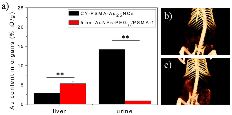
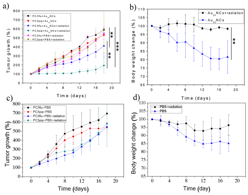
For over a hundred years, X-rays have been a main component of the radiotherapeutic approaches to treat cancer. Yet, to date, no radiosensitizer has been developed to selectively target prostate cancer. Gold has excellent X-ray absorptivity and is used as a radiotherapy enhancing material. In this work, ultrasmall Au25 nanoclusters (NCs) are developed for selective prostate cancer targeting, radiotherapy enhancement, and rapid clearance from the body. Targeted-Au25 NCs are rapidly and selectively taken up by prostate cancer in vitro and in vivo and also have fast renal clearance. When combined with X-ray irradiation of the targeted cancer tissues, radiotherapy is significantly enhanced. The selective targeting and rapid clearance of the nanoclusters may allow reductions in radiation dose, decreasing exposure to healthy tissue and making them highly attractive for clinical translation.

Keywords: PSMA; gold nanoclusters; prostate cancer; radiosensitization; targeted radiotherapy.


Conflict of interest statement

Conflict of Interest

The authors declare no conflict of interest.

Figures

Figure 1.
Figure 1.
CY-PSMA-1-Au25NCs for targeted radiotherapy of prostate cancer. a) Schematic of synthesis of CY-PSMA-1-Au25NCs radiosensitizer, with high affinity to the PSMA-expressing PC3pip cells. b) Illustration of PC3pip and PC3flu tumor targeting and radiation therapy with CY-PSMA-1-Au25NCs. PSMA-expressing PC3pip cells and PSMA-negative PC3flu cells are implanted on the flanks of the same mouse. Intravenous injection of nanoclusters results in higher targeting levels to the PC3pip tumor than to the PC3flu tumor and, thus, improved radiation therapy.
Figure 2.
Figure 2.
a) Distribution of core diameters of CY-PSMA-1-Au25NCs. Inset shows a TEM image of Au25NCs (scale bar 5 nm). b) Distribution of the hydrodynamic diameters of CY-PSMA-1-Au25NCs. c) Excitation and emission spectra of nanoclusters showing peaks at 490 nm (ex) and 700 nm (em). d) MALDI-MS spectra for CY-PSMA-1-Au25NCs. A series of intense peaks between 5–12k m/z fit the formula Au25S(18-m)Pm (P=CY-PSMA-1) (m= 0–18), and the difference between adjacent intense peaks is 1320, which matches the molecular weight of CY-PSMA-1 with one thiol group missing. The inset is an expanded view of peak details in the range 6.2k-7k. Space between the minor peaks is 197 m/z and 32 m/z, which corresponds to the loss of Au and S atoms, respectively. Matrix, CHCA, linear model.
Figure 3.
Figure 3.
a) Confocal images of the uptake of CY-PSMA-1-Au25NCs by PC3pip and PC3flu cells after 24 h incubation at 30 μg Au/mL. Au25NCs show a red fluorescence and DAPI is used for nuclear staining (scale bar 25 μm). b) ICP-MS measurement of Au content in cells, showing significantly higher Au content in PC3pip cells than in PC3flu cells. c) Competition binding curves for parent ZJ24, CY-PSMA-1 ligands, and CY-PSMA-1-Au25NCs. d) In vitro ROS generation by a DCFDA assay. Non-fluorescent DCFDA was converted into fluorescent DCF by intracellular ROS upon 6 Gy radiation. e) Survival curves of PC3pip and PC3flu cells with and without addition of CY-PSMA-1-Au25NCs under radiation at doses of 0, 2, 4, and 6 Gy. Data are presented as mean ± SD. n = 3; two-tailed t-test: **P < 0.05.
Figure 4.
Figure 4.
a) In vivo 3D CT images of the PC3pip (right, blue) and PC3flu (left, green) tumor-bearing mice (indicated by blue and green ovals) before and at 0.5 h, 1 h, 2 h, 4 h, 6 h, and 24 h after intravenous injection of CY-PSMA-1-Au25NCs (30 μg Au/g mouse); b) The CT signals at the tumor regions and bladder at each time point after CY-PSMA-1-Au25NCs injection; c) In vivo 3D CT images of the bladders of mice (indicated by red circles) at each time point; d) Biodistribution of CY-PSMA-1-Au25NCs in the main organs/urine at 4 h and 24 h post injection, as determined by ICP-MS (data are presented as mean ± SD. n = 3; two-tailed t-test: **P < 0.05).
Figure 5.
Figure 5.
a) Au content in liver and urine of mice injected with CY-PSMA-1-Au25NCs and 5 nm AuNPs-PEG2K/PSMA-1 at 24 h post injection, as determined by ICP-MS (data are presented as mean ± SD. n = 3; two-tailed t-test: **P < 0.05); In vivo 3D CT images of the digestive system of mice injected with b) CY-PSMA-1-Au25NCs and c) 5 nm AuNPs-PEG2K/PSMA-1 at 24 h post injection, showing more accumulation of 5 nm AuNPs-PEG2K/PSMA-1 in the gut than that measured for CY-PSMA-1-Au25NCs.
Figure 6.
Figure 6.
Tumor growth curves and body weight changes of mice after treated with a, b) CY-PSMA-1-Au25NCs only and both CY-PSMA-1-Au25NCs and 6 Gy radiation, and c, d) PBS only and both PBS and 6 Gy radation. For comparison, tumor growth curves of mice injected with PBS and radiation were also plotted in a). (data are presented as mean ± SD. n = 3; two-tailed t-test: **p≤0.05, ***p≤0.01).

References

    1. Siegel RL, Miller KD, Jemal A, CA Cancer J. Clin 2018, 68, 7. - PubMed
    1. Wang X, Tsui B, Ramamurthy G, Zhang P, Meyers J, Kenney ME, Kiechle J, Ponsky L, Basilion JP, Mol. Cancer Ther 2016, 15, 1834. - PubMed
    1. Zhu S, Gu Z, Zhao Y, Adv. Ther 2018, 1, 1800050.
    1. Song G, Cheng L, Chao Y, Yang K, Liu Z, Adv. Mater 2017, 29, 1700996. - PubMed
    1. Hainfeld JF, Dilmanian FA, Slatkin DN, Smilowitz HM, J. Pharm. Pharmacol 2008, 60, 977. - PubMed

Publication types