. 2019 Oct:193:105422.
doi: 10.1016/j.jsbmb.2019.105422. Epub 2019 Jun 29.

DLK1/PREF1 marks a novel cell population in the human adrenal cortex

Affiliations

DLK1/PREF1 marks a novel cell population in the human adrenal cortex

Irene Hadjidemetriou et al. J Steroid Biochem Mol Biol. 2019 Oct.

Abstract

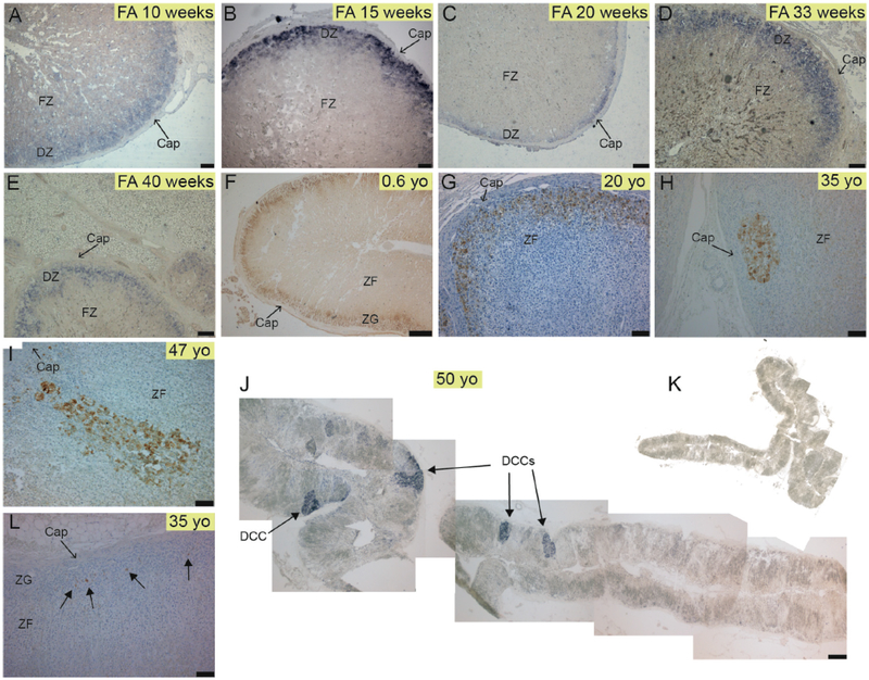
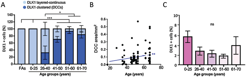
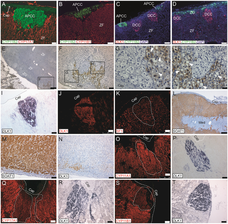
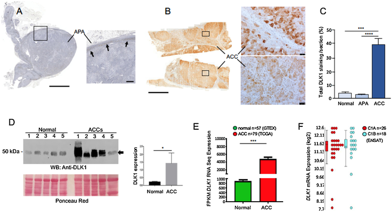
The adrenal cortex governs fundamental metabolic processes though synthesis of glucocorticoid, mineralocorticoids and androgens. Studies in rodents have demonstrated that the cortex undergoes a self-renewal process and that capsular/subcapsular stem/progenitor cell pools differentiate towards functional steroidogenic cells supporting the dynamic centripetal streaming of adrenocortical cells throughout life. We previously demonstrated that the Notch atypical ligand Delta-like homologue 1 (DLK1)/preadipocyte factor 1 (PREF1) is expressed in subcapsular Sf1 and Shh-positive, CYP11B1-negative and CYP11B2-partially positive cortical progenitor cells in rat adrenals, and that secreted DLK1 can modulate GLI1 expression in H295R cells. Here we show that the human adrenal cortex remodels with age to generate clusters of relatively undifferentiated cells expressing DLK1. These clusters (named DLK1-expressing cell clusters or DCCs) increased with age in size and were found to be different entities to aldosterone-producing cell clusters, another well-characterized and age-dependent cluster structure. DLK1 was markedly overexpressed in adrenocortical carcinomas but not in aldosterone-producing adenomas. Thus, this data identifies a novel cell population in the human adrenal cortex and might suggest a yet-to be identified role of DLK1 in the pathogenesis of adrenocortical carcinoma in humans.

Keywords: Adrenal cortex; Adrenocortical carcinoma; DLK1; Remodeling; Steroidogenesis.


Conflict of interest statement

Declaration of competing interests

The author declare no conflict of interest.

Figures

Fig. 1.
Fig. 1.. The human adrenal cortex remodels to generate clusters of cells expressing DLK1.
A-E) Expression of DLK1 mRNA in human fetal adrenals showing a subcapsular layered-continuous pattern in the definitive zone (DZ). F-G) DLK1 protein expression in a 0.6 yo and 20 yo adrenal showing a subcapsular layered-continuous pattern. H) A round-shaped subcapsular DCC in a 35 yo adrenal. I) A columnar-shaped DCC in a 47 yo adrenal. J) Expression of DLK1 mRNA in a 50 yo adrenal showing multiple DCCs and absence of a layered-continuous pattern of DLK1 expression. K) Control sense probe in an adjacent section to J. L) From the third decade onwards, large areas of the cortex were devoid of either clustered or layered-continuous DLK1 staining, and have single cells sparingly expressing DLK1 or no DLK1 expression at all. Abbreviations: Cap, capsule; DCC, DLK1 cells cluster; DZ, Definitive Zone; FZ, fetal Zone; ZG, Zona Glomerulosa; ZF, Zona Fasciculata. Scale bars A, B = 75 μm; C = 200 μm; D, E, G, H, I, L = 100 μm; F = 500 μm; J = 1500 μm. Hadjidemetriou et al., Fig 2 (1.5 columns fitting).
Fig. 2.
Fig. 2.. DCCs appear in the third decade of life and increase in size with age.
A) Clustered (DCCs) and layered-continuous DLK1 expression in physiologically normal human adrenals are expressed as percentage of total cortical DLK1 staining in each age group. Error bars represent standard error of the mean, two-way ANOVA: * (p < 0.05), *** (p < 0.001) B) Quantitative analysis of the size of DCCs with age. Pearson correlation test. C) Percentage of total DLK1 staining expressed as percentage of DLK1 staining/total cortical area showing a non-significant trend of reduction of DLK1 positivity up to approximately 60 yo. ns, not significant. For A–C (also applicable to Fig. 1), n = 11 (FAs), 7 (0–25 yo), 6 (26–40 yo), 5 (41–50 yo), 6 (51–60 yo), 8 (61–70 yo). Hadjidemetriou et al., Fig 3 (2 columns fitting).
Fig. 3.
Fig. 3.. DCCs are different entities to APCCs and are incompletely differentiated.
A–B) APCCs of different shapes and sizes can be detected by specific anti-CYP11B2 antibodies and were subcapsular in CYP17A1 and CYP11B1 negative cells. C) Double staining with DLK1 and CYP11B2 showing that DCCs and APCCs are different cellular entities in the adrenal cortex. Two APCCs and two DCCs are seen. D) DCCs can be located adjacent to CYP11B2 positive cells with the latter organized in a classical ZG manner (subcapsular layered-continuous); even in this case double-positive cells are not visible. Age of adrenal in A–D is 50 yo. E–H) A DCC in the adrenal of a 28 yo donor stained with anti DLK1 antibodies and counterstained with hematoxylin. Structural features of DCC are in G and H, where vacuoles are indicated by white arrowheads. I–J) Three consecutive sections were processes for DLK1 in situ hybridization (I) or DLK1 IF (J), showing an identical pattern of DLK1 mRNA and protein expression. The third section was stained with anti SF1 antibodies (K), showing DCCs to be SF1 positive. L–N) SOAT1 is expressed throughout the cortex but not in the capsule and in the medulla (L). Consecutive adrenal sections were processes for SOAT1 (M) and DLK1 (N) IHC showing DCCs to be SOAT1 positive. O–P) Consecutive adrenal sections were processed for CYP11A1 IF (O) and DLK1 in situ hybridization (P), showing DCCs to be CYP11A1 positive. Q–R) Consecutive adrenal sections were processes for CYP17A1 IF (Q) and DLK1 in situ hybridization (R), showing DCCs to express low/nil levels of CYP17A1. S–T) Consecutive adrenal sections were processes for CYP11B1 IF (S) and DLK1 in situ hybridization (T), showing DCCs to express low/nil levels of be CYP11B1. Abbreviations: APCC: Aldosterone-Producing Cell Cluster; Cap, capsule; DCCs, DLK1 Cells Clusters; ZG, Zona Glomerulosa; ZF, Zona Fasciculata; Med., medulla. Scale bars A–D = 75 μm; E and L = 500 μm; F, I–K and M–T = 100 μm; G, H = 25 μm. n = 3 or > 3 each experiment. Hadjidemetriou et al., Fig 4 (2 columns fitting).
Fig. 4.
Fig. 4.. DLK1 is overexpressed in adrenocortical carcinomas.
Immunohistochemical detection of DLK1 protein in APA (A) and ACC sections (cohort 1, B). In APAs, detectable levels of DLK1 staining are seen in the subcapsular region only (arrows). Note the widespread DLK1 staining in ACC compared to APA. However, in each section analyzed, DLK1 staining was not homogeneous within the ACC parenchyma, with areas of high (top right panel in B) and low (bottom right panel in B) DLK1 expression. C) Quantification of DLK1 staining in ACC, APA and normal adrenal sections (cohort 1) showed a significant higher expression of DLK1 in ACC compared to both normal adrenals and APAs. No ENSAT/Weiss scoring as well as hormonal status was available for these samples. D) Western blot analysis of DLK1 expression in 5 normal adrenals and 5 ACC lysates (cohort 2) showing up-regulation of DLK1 (arrow) in ACC samples. ACCs were as follow: 1) score 2 (ENSAT), score 4/9 (Weiss), cortisol secreting; 2) score 2 (ENSAT), score 6/9 (Weiss), no secreting; 3) score 3 (ENSAT), score 8/9 (Weiss), androgens secreting (plus cortisol from urine steroid profiling); 4) score 3 (ENSAT), score 9/9 (Weiss), androgens and cortisol secreting; 5) score 2 (ENSAT), score 8/9 (Weiss), androgens secreting (plus cortisol from urine steroid profiling). Densitometric analysis of western blot bands is provided in the inset. E) DLK1 transcript expression is significantly higher in ACC compared to normal adrenal, ***(p < 0.001). Green – normal adrenal transcript GTeX (n = 45), Red – ACC transcript TCGA (n = 79) F) DLK1 level is consistently upregulated on mRNA level using ENSAT microarray gene expression data. Error bars in C represent +/− SEM; One-way ANOVA, with statistical significance denoted as ***(p < 0.001) and ****(p < 0.0001). Error bars in panel to D represent +/− SEM; Mann–Whitney–Wilcoxon test, *(p < 0.05). Analysis of E and F as in Methods. Scale bars A and B = 5000 μm; inset to A = 500 μm; insets to B = 50 μm.

References

    1. King P, Paul A, Laufer E, Shh signaling regulates adrenocortical development and identifies progenitors of steroidogenic lineages, Proc. Natl. Acad. Sci. U. S. A 106 (50) (2009) 21185–21190. - PMC - PubMed
    1. Guasti L, et al., Dlk1 up-regulates Gli1 expression in male rat adrenal capsule cells through the activation of beta1 integrin and ERK1/2, Endocrinology 154 (12) (2013) 4675–4684. - PubMed
    1. Finco I, Lerario AM, Hammer GD, Sonic hedgehog and WNT signaling promote adrenal gland regeneration in male mice, Endocrinology 159 (2) (2018) 579–596. - PMC - PubMed
    1. Falix FA, et al., Possible roles of DLK1 in the Notch pathway during development and disease, Biochim. Biophys. Acta 1822 (6) (2012) 988–995. - PubMed
    1. Floridon C, et al., Does fetal antigen 1 (FA1) identify cells with regenerative, endocrine and neuroendocrine potentials? a study of FA1 in embryonic, fetal, and placental tissue and in maternal circulation, Differentiation 66 (1) (2000) 49–59. - PubMed

Publication types

MeSH terms

LinkOut - more resources