

T cell-selective deletion of Oct1 protects animals from autoimmune neuroinflammation while maintaining neurotropic pathogen response
- PMID: 31266507
- PMCID: PMC6607600
- DOI: 10.1186/s12974-019-1523-3
T cell-selective deletion of Oct1 protects animals from autoimmune neuroinflammation while maintaining neurotropic pathogen response
Abstract
Background: Treatments for autoimmune diseases aim to dampen autoreactivity while preserving normal immune function. In CD4+ T cells, the transcription factor Oct1/Pou2f1 is a dispensable transcription factor for T cell development and response to primary infection, but promotes expression of target genes, including Il2 and Ifng, under conditions of antigen reencounter. As a result, they are more strongly expressed upon secondary stimulation. Such repeated antigen encounters occur in memory recall responses, in autoimmunity where self-antigen can be recognized multiple times, and in chronic infection where foreign antigen is persistent. Based on these previous findings, we hypothesized that Oct1 loss would protect animals from autoimmunity but maintain normal responses to pathogens in the CNS.
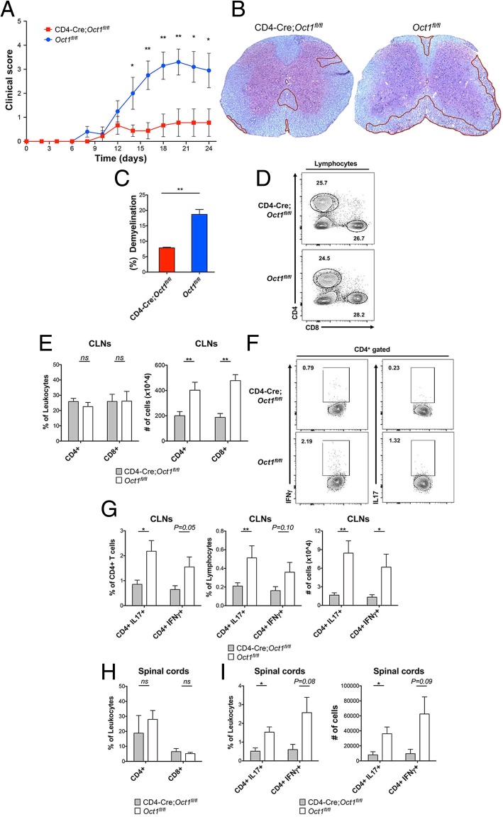
Objective: We used a conditional mouse Oct1 (Pou2f1) allele and a CD4-Cre driver to determine the effect of T cell-specific Oct1 loss on autoimmune- and viral-induced neuroinflammation using an autoantigen-driven EAE model of autoimmunity and a JHMV model of viral infection.
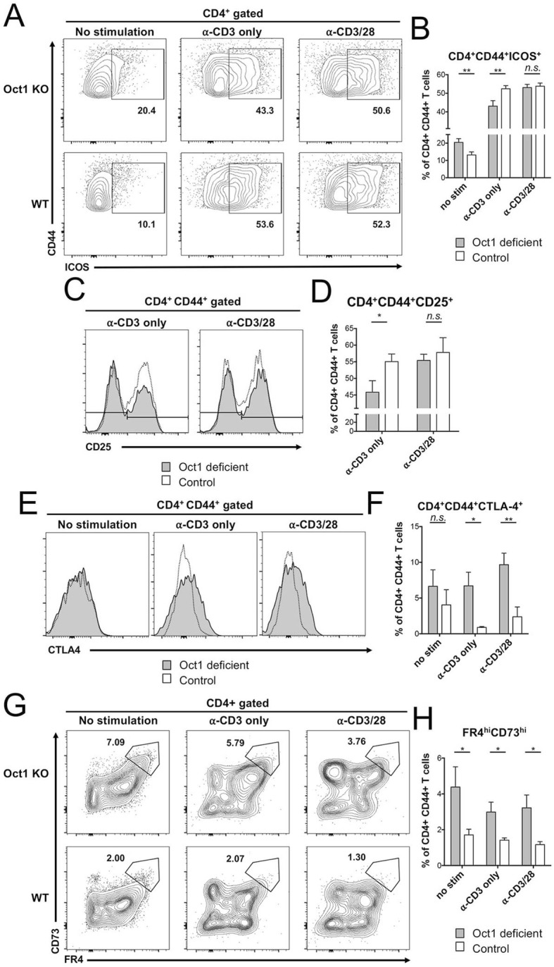
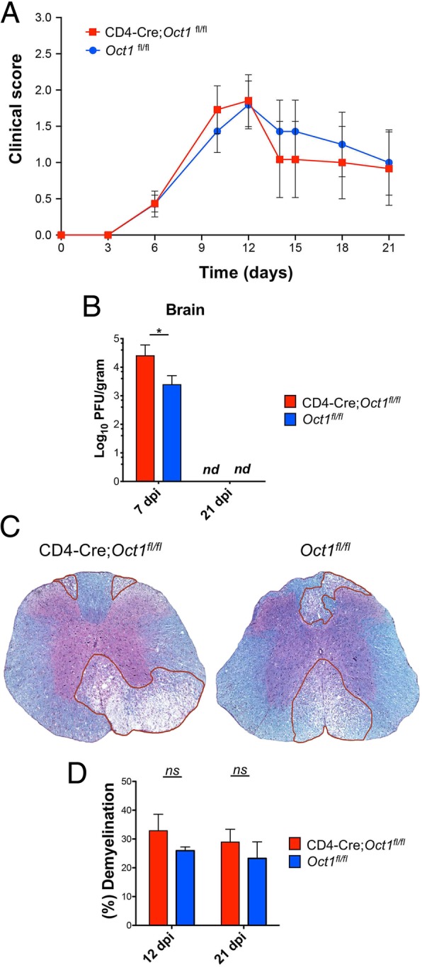
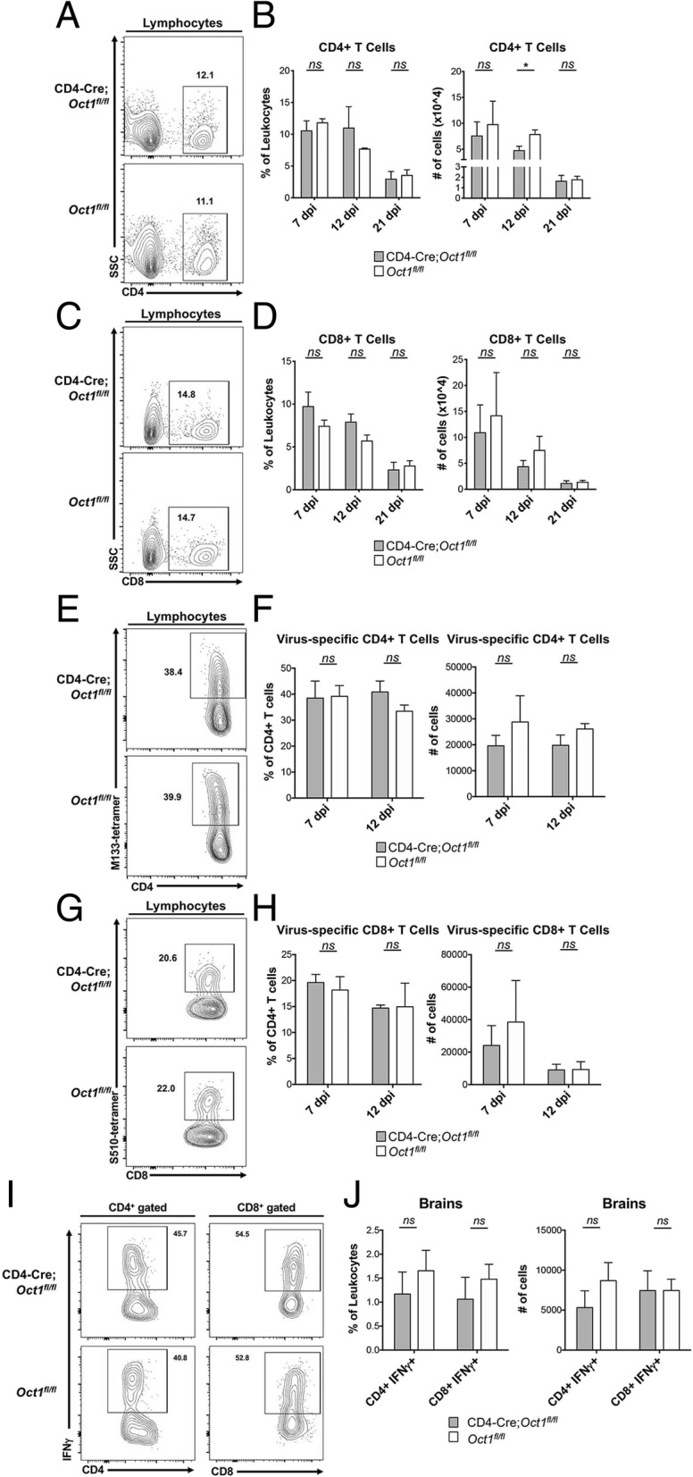
Results: Oct1 conditional deletion mitigated clinical scores and reduced infiltrating T cells and cytokine production in the EAE model. Consistently, Oct1-deficient CD4+ T cells stimulated in vitro showed increased expression of markers associated with T cell anergy, particularly in the absence of co-stimulatory signals. In contrast, anti-viral T cell effector functions are intact in the absence of Oct1, with no changes in neuroinflammation, infiltrating T cells or cytokine production.
Conclusion: Our findings uncover a significant difference between the effect of Oct1 loss on autoimmune and anti-pathogen responses, which potentially could be exploited for therapeutic benefit.
Keywords: Experimental autoimmune encephalomyelitis; JHMV; Oct1/POU2F1; T lymphocytes.
Conflict of interest statement
The authors declare that they have no competing interests.
Figures
Similar articles
-
Dopaminergic stimulation leads B-cell infiltration into the central nervous system upon autoimmunity.J Neuroinflammation. 2021 Dec 17;18(1):292. doi: 10.1186/s12974-021-02338-1. J Neuroinflammation. 2021. PMID: 34920747 Free PMC article.
-
Oct1 and OCA-B are selectively required for CD4 memory T cell function.J Exp Med. 2015 Nov 16;212(12):2115-31. doi: 10.1084/jem.20150363. Epub 2015 Oct 19. J Exp Med. 2015. PMID: 26481684 Free PMC article.
-
Regulation of autoreactive CD4 T cells by FoxO1 signaling in CNS autoimmunity.J Neuroimmunol. 2021 Oct 15;359:577675. doi: 10.1016/j.jneuroim.2021.577675. Epub 2021 Jul 31. J Neuroimmunol. 2021. PMID: 34403862 Free PMC article.
-
Therapeutic benefits of regulating inflammation in autoimmunity.Inflamm Allergy Drug Targets. 2008 Sep;7(3):203-10. doi: 10.2174/187152808785748155. Inflamm Allergy Drug Targets. 2008. PMID: 18782028 Review.
-
The Oct1 transcription factor and epithelial malignancies: Old protein learns new tricks.Biochim Biophys Acta. 2016 Jun;1859(6):792-804. doi: 10.1016/j.bbagrm.2016.02.007. Epub 2016 Feb 10. Biochim Biophys Acta. 2016. PMID: 26877236 Free PMC article. Review.
Cited by
-
Impact of preweaning vaccination on host gene expression and antibody titers in healthy beef calves.Front Vet Sci. 2022 Sep 26;9:1010039. doi: 10.3389/fvets.2022.1010039. eCollection 2022. Front Vet Sci. 2022. PMID: 36225796 Free PMC article.
-
HSV-1 selectively packs the transcription factor Oct-1 into EVs to facilitate its infection.Front Microbiol. 2023 Jun 15;14:1205906. doi: 10.3389/fmicb.2023.1205906. eCollection 2023. Front Microbiol. 2023. PMID: 37396389 Free PMC article.
-
OCA-B promotes pathogenic maturation of stem-like CD4+ T cells and autoimmune demyelination.J Clin Invest. 2025 Apr 29;135(13):e187862. doi: 10.1172/JCI187862. eCollection 2025 Jul 1. J Clin Invest. 2025. PMID: 40299553 Free PMC article.
-
Targeting transcriptional coregulator OCA-B/Pou2af1 blocks activated autoreactive T cells in the pancreas and type 1 diabetes.J Exp Med. 2021 Mar 1;218(3):e20200533. doi: 10.1084/jem.20200533. J Exp Med. 2021. PMID: 33295943 Free PMC article.
-
POU2F1 Promotes Cell Viability and Tumor Growth in Gastric Cancer through Transcriptional Activation of lncRNA TTC3-AS1.J Oncol. 2021 Jun 28;2021:5570088. doi: 10.1155/2021/5570088. eCollection 2021. J Oncol. 2021. PMID: 34257651 Free PMC article.
References
MeSH terms
Substances
Grants and funding
LinkOut - more resources
Full Text Sources
Molecular Biology Databases
Research Materials
