. 2019 Aug 23;294(34):12670-12682.
doi: 10.1074/jbc.RA119.009368. Epub 2019 Jul 2.

Two complementary α-fucosidases from Streptococcus pneumoniae promote complete degradation of host-derived carbohydrate antigens

Affiliations

Two complementary α-fucosidases from Streptococcus pneumoniae promote complete degradation of host-derived carbohydrate antigens

Joanne K Hobbs et al. J Biol Chem. .

Abstract

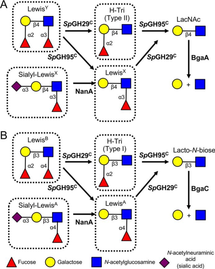
An important aspect of the interaction between the opportunistic bacterial pathogen Streptococcus pneumoniae and its human host is its ability to harvest host glycans. The pneumococcus can degrade a variety of complex glycans, including N- and O-linked glycans, glycosaminoglycans, and carbohydrate antigens, an ability that is tightly linked to the virulence of S. pneumoniae Although S. pneumoniae is known to use a sophisticated enzyme machinery to attack the human glycome, how it copes with fucosylated glycans, which are primarily histo-blood group antigens, is largely unknown. Here, we identified two pneumococcal enzymes, SpGH29C and SpGH95C, that target α-(1→3/4) and α-(1→2) fucosidic linkages, respectively. X-ray crystallography studies combined with functional assays revealed that SpGH29C is specific for the LewisA and LewisX antigen motifs and that SpGH95C is specific for the H(O)-antigen motif. Together, these enzymes could defucosylate LewisY and LewisB antigens in a complementary fashion. In vitro reconstruction of glycan degradation cascades disclosed that the individual or combined activities of these enzymes expose the underlying glycan structure, promoting the complete deconstruction of a glycan that would otherwise be resistant to pneumococcal enzymes. These experiments expand our understanding of the extensive capacity of S. pneumoniae to process host glycans and the likely roles of α-fucosidases in this. Overall, given the importance of enzymes that initiate glycan breakdown in pneumococcal virulence, such as the neuraminidase NanA and the mannosidase SpGH92, we anticipate that the α-fucosidases identified here will be important factors in developing more refined models of the S. pneumoniae-host interaction.

Keywords: Streptococcus; X-ray crystallography; glycoside hydrolase; host-pathogen interaction; structure-function.


Conflict of interest statement

The authors declare that they have no conflicts of interest with the contents of this article

Figures

Figure 1.
Figure 1.
Overall structure of SpGH29C. The X-ray crystal structure of SpGH29C is represented as a cartoon colored from blue (N terminus (Nter)) to red (C terminus (Cter)). Both catalytic residues and a Bis-Tris molecule observed to be bound in the active site are shown as gray sticks. The numbering of helices and β-strands comprising the (α/β)8 catalytic module is indicated. The strand numbering of the ancillary module is also indicated.
Figure 2.
Figure 2.
The SpGH29CT D171N/E215Q catalytic pocket in complex with histo-blood group antigens. A, structure of SpGH29T D171N/E215Q (green sticks) in complex with LewisA antigen. The insets focus on the +2′ galactose-binding subsite (top right inset), the −1 fucose-binding subsite (bottom right inset), and +1* GlcNAc pseudo-subsite (left inset). B, structural overlay of BiAfcB D172A/E217A (PDB code 3EUT; orange sticks) in complex with the LewisA antigen (gray lines) with the LewisA antigen from the complex with SpGH29T D171N/E215Q. The active-site side chains of SpGH29T D171N/E215Q were omitted as they are completely conserved with those of BiAfcB. SpGH29T residue numbering is shown in black, and that of BiAfcB is shown in gray. C and D, SpGH29T D171N/E215Q in complex with the LewisX and LewisY antigens, respectively. Fucose, galactose, and GlcNAc are shown in red, yellow, and blue sticks, respectively. The water molecule (wat) is represented by a purple sphere. Dashed lines denote hydrogen bonds. Subsites are indicated in red.
Figure 3.
Figure 3.
Example of sequential degradation of a human glycan by pneumococcal GHs. A, the substrate type IV H-tetrasaccharide was incubated with enzyme(s) overnight, and the products were labeled and visualized by fluorophore-assisted carbohydrate electrophoresis. ± above the gel indicates the presence/absence of substrate or enzyme. B, schematic depiction of the sequential breakdown of type IV H-tetrasaccharide by pneumococcal GHs. GHs are indicated in bold next to the arrow for the reaction they catalyze.
Figure 4.
Figure 4.
Schematic depiction of the sequential degradation of histo-blood group antigens by pneumococcal GHs. GHs are indicated in bold next to the arrow for the reaction they catalyze. A, degradation of LewisY can be initiated either by SpGH29C, which yields the type II H-trisaccharide (H-Tri), or by SpGH95C, which yields LewisX. The complementary α-fucosidase then acts to produce N-acetyllactosamine (LacNAc), which is cleaved into its constituent monosaccharides by BgaA. Sialyl-LewisX must be desialylated by NanA prior to SpGH29C activity. B, degradation of LewisB can be initiated either by SpGH29C, which yields the type I H-trisaccharide, or by SpGH95C, which yields LewisA. The complementary α-fucosidase then acts to produce lacto-N-biose, which is cleaved into its constituent monosaccharides by BgaC. Sialyl-LewisA must be desialylated by NanA prior to SpGH29C activity. See Fig. S3 for experimental validation for the sequential depolymerization of each of the boxed species in this figure.
Figure 5.
Figure 5.
Cellular localization of SpGH29C. A and B, activity of different cellular fractions of WT TIGR4 (A) and ΔspGH29C (B) against LewisX as detected by fluorophore-assisted carbohydrate electrophoresis. The activities of recombinant SpGH29C and BgaA against LewisX are shown as controls, and fucose is shown as a standard. C, background labeling of the different cellular fractions in the absence of LewisX. LewisX and LewisX treated with total soluble protein are shown for comparison. LeX, LewisX; EF, extracellular fraction; CF, cytoplasmic fraction; MF, membrane fraction. The 8-aminonaphthalene-1,3,6-trisulfonic acid (ANTS) lane indicates background labeling due to the fluorophore alone. Due to the background labeling of the cell wall–associated fraction, SpGH29C activity can be observed as a disappearance of LewisX rather than an appearance of fucose.
Figure 6.
Figure 6.
Schematic depiction of the sequential degradation of TFLNH by pneumococcal GHs. GHs are indicated in bold next to the arrow for the reaction they catalyze; numbers in green refer to the gel lane in Fig. S4. SpGH95C and SpGH29C are required to remove the capping fucose residues from TFLNH and allow access to the oligosaccharide by other GHs. Treatment of TFLNH with SpGH29C results in removal of the α-(1→3)– and α-(1→4)–linked fucose units and allows BgaA and GH20C to degrade the arm proximal to the reducing end; however, without SpGH95C, the distal arm cannot be degraded. Treatment of TFLNH with SpGH95C results in partial removal of the α-(1→2)–linked fucose unit and a difucosylated oligosaccharide that cannot be acted upon by other GHs (except SpGH29C). If the α-(1→3)– and α-(1→4)–linked fucose units are removed by SpGH29C first, SpGH95C is able to fully remove the α-(1→2)–linked fucose unit from the distal arm to produce lacto-N-hexaose. This hexasaccharide can then be fully degraded into galactose and glucose by the combined actions of BgaA, BgaC, and GH20C. See Fig. S4 for experimental validation of this pathway.

References

    1. Varki A., and Marth J. (1995) Oligosaccharides in vertebrate development. Semin. Dev. Biol. 6, 127–138 10.1016/S1044-5781(06)80022-8 - DOI
    1. Khoury G. A., Baliban R. C., and Floudas C. A. (2011) Proteome-wide post-translational modification statistics: frequency analysis and curation of the Swiss-Prot database. Sci. Rep. 1, srep00090 10.1038/srep00090 - DOI - PMC - PubMed
    1. Gagneux P., Aebi M., and Varki A. (2015) Evolution of glycan diversity, in Essentials of Glycobiology (Varki A., Cummings R. D., Esko J. D., Stanley P., Hart G. W., Aebi M., Darvill A. G., Kinoshita T., Packer N. H., Prestegard J. H., Schnaar R. L., and Seeberger P. H., eds) 3rd Ed., Cold Spring Harbor Laboratory, Cold Spring Harbor, NY - PubMed
    1. Reitsma S., Slaaf D. W., Vink H., van Zandvoort M. A., and oude Egbrink M. G. (2007) The endothelial glycocalyx: composition, functions, and visualization. Pflugers Arch. 454, 345–359 10.1007/s00424-007-0212-8 - DOI - PMC - PubMed
    1. Unione L., Gimeno A., Valverde P., Calloni I., Coelho H., Mirabella S., Poveda A., Arda A., and Jimenez-Barbero J. (2017) Glycans in infectious diseases. A molecular recognition perspective. Curr. Med. Chem. 24, 4057–4080 10.2174/0929867324666170217093702 - DOI - PubMed

Publication types

MeSH terms

LinkOut - more resources